. 2005 Mar;25(6):2511-24.
doi: 10.1128/MCB.25.6.2511-2524.2005.

Human Fas-associated factor 1, interacting with ubiquitinated proteins and valosin-containing protein, is involved in the ubiquitin-proteasome pathway

Affiliations

Human Fas-associated factor 1, interacting with ubiquitinated proteins and valosin-containing protein, is involved in the ubiquitin-proteasome pathway

Eun Joo Song et al. Mol Cell Biol. 2005 Mar.

Abstract

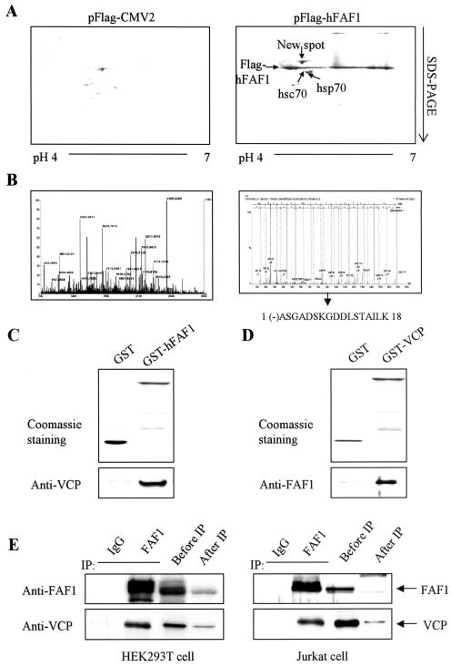
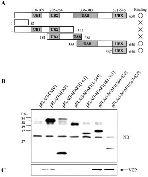
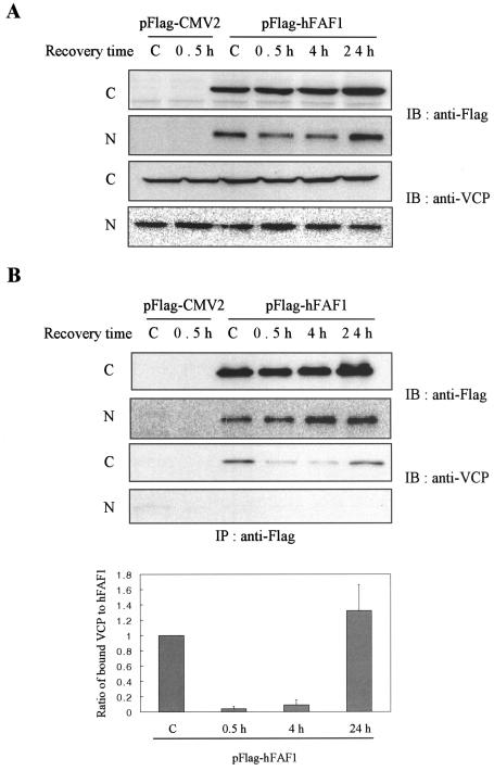
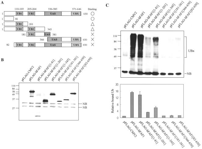
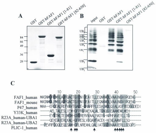
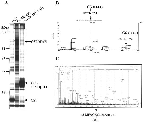
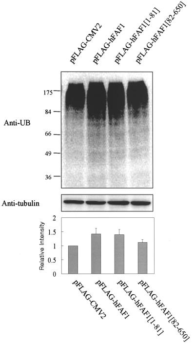
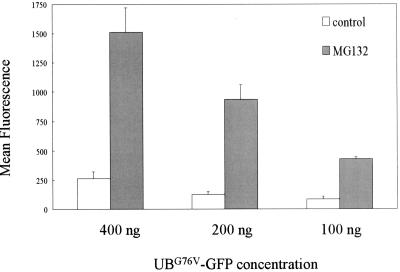
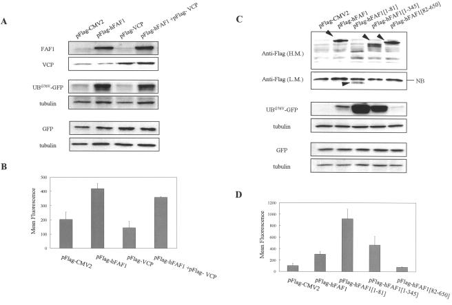
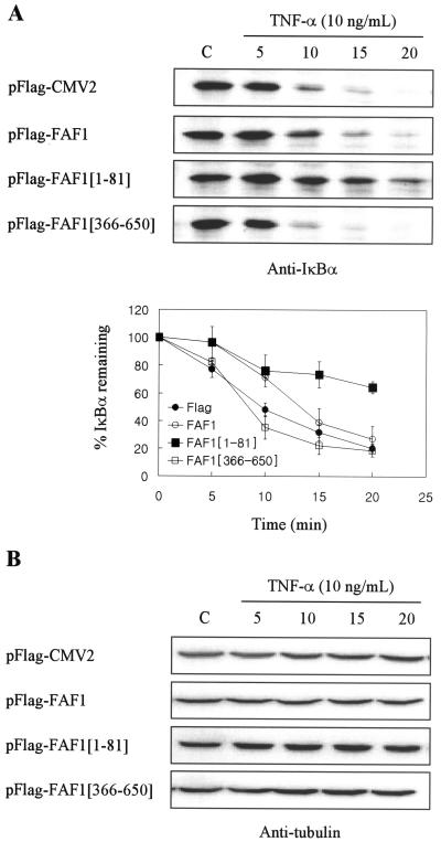
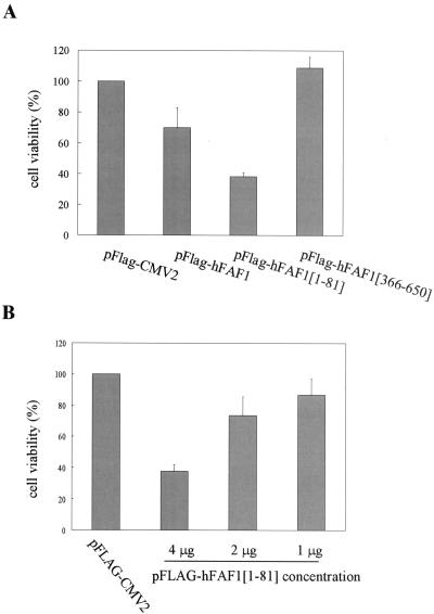
Human Fas-associated factor 1 (hFAF1) is a novel protein having multiubiquitin-related domains. We investigated the cellular functions of hFAF1 and found that valosin-containing protein (VCP), the multiubiquitin chain-targeting factor in the degradation of the ubiquitin-proteasome pathway, is a binding partner of hFAF1. hFAF1 is associated with the ubiquitinated proteins via the newly identified N-terminal UBA domain and with VCP via the C-terminal UBX domain. The overexpression of hFAF1 and a truncated UBA domain inhibited the degradation of ubiquitinated proteins and increased cell death. These results suggest that hFAF1 binding to ubiquitinated protein and VCP is involved in the ubiquitin-proteasome pathway. We hypothesize that hFAF1 may serve as a scaffolding protein that regulates protein degradation in the ubiquitin-proteasome pathway.


Figures

FIG. 1.
FIG. 1.
hFAF1 interacts with VCP in vitro and in vivo. (A) HEK293T cells transfected with pFlag-CMV-2 vector and pFlag-hFAF1 were labeled with 1 μCi of [35S]methionine/ml. Immunoprecipitation in cell lysates was carried out with anti-Flag M2 agarose cross-linking affinity beads (Sigma). Precipitates were analyzed by two-dimensional gel electrophoresis and autoradiographed with BAS2500. (B) Protein spots detected in the autoradiograph were cut out from the corresponding silver-stained gel and subjected to in-gel digestion with trypsin. Mass peptide fingerprint analyses were conducted by MALDI-TOF MS (left spectrum), and peptides were sequenced by using ESI-q-TOF tandem MS (right spectrum). (C and D) GST-hFAF1 (C) and GST-VCP (D) were immobilized on glutathione beads and incubated with purified recombinant VCP (C) and purified recombinant hFAF1 (D). Binding proteins were detected on SDS-PAGE with Coomassie staining and immunoblotted by anti-VCP antibody (C) and anti-FAF1 antibody (D). (E) For the interaction between endogenous FAF1 and VCP, HEK293T cells and Jurkat cell lysates were immunoprecipitated with mouse immunoglobulin G control (lane 1) and anti-FAF1 polyclonal antibodies (lane 2). The amount of endogenous FAF1 and VCP in 10% of cell lysates before immunoprecipitation was compared with the amounts after immunoprecipitation. Immunoprecipitates were analyzed by Western blotting using anti-FAF1 and anti-VCP antibodies.
FIG. 2.
FIG. 2.
hFAF1 interacts with VCP via the UBX domain. (A) Diagram of various truncated forms of Flag-hFAF1. UB1 and UB2, ubiquitin homologous domains; UAS domain, homologous with Caenorhabditis elegans open reading frame C281.1; UBX, domain present in ubiquitin regulatory proteins. (B and C) HEK293T cells transfected with pFlag-CMV-2 vector or truncated Flag-hFAF1 were lysed and immunoprecipitated with monoclonal anti-Flag M2 agarose cross-linking affinity gel (Sigma). Precipitates were analyzed by Western blotting using anti-Flag (B) and anti-VCP (C) antibodies. NB indicates a nonspecific protein band.
FIG. 3.
FIG. 3.
hFAF1 interacts with VCP in cytosol. HEK293T cells transiently transfected with pFlag-CMV-2 vector or Flag-hFAF1 were heat shocked at 45°C for 45 min and recovered for the indicated times. “C” lane headings indicate untreated cells. At each time point, cells were fractionated into cytosolic (C) and nuclear (N) fractions and Western blotted with anti-Flag and anti-VCP antibodies (A). For examining the binding, each fraction was immunoprecipitated with monoclonal anti-Flag M2 agarose cross-linking affinity gel, and precipitates were analyzed by Western blot analysis using anti-Flag and anti-VCP antibodies. Western analysis of panel B was quantified with ImageQuant (Amersham Biosciences). The relative ratio of bound VCP to Flag-hFAF1 at control is arbitrarily presented as 1. Bars show means ± standard deviations (SD) from three independent experiments (B).
FIG. 4.
FIG. 4.
hFAF1 binds to multiubiquitinated proteins in vivo. (A) Diagram of various truncate forms of Flag-hFAF1. (B and C) HEK293T cells transfected with pFlag-CMV-2 vector or truncated forms of Flag-hFAF1 were lysed and immunoprecipitated with monoclonal anti-Flag M2 agarose cross-linking affinity gel (Sigma). Precipitates were analyzed by Western blotting using anti-Flag (B) and antiubiquitin (C) antibodies. NB indicates a nonspecific band, and UBn indicates multiubiquitinated proteins. Western analysis of panel C was quantified with ImageQuant (Amersham Biosciences). The relative ratio of bound multiubiquitinated proteins to pFlag-CMV2 is arbitrarily presented as 1. Bars show means ± SD from three independent experiments.
FIG. 5.
FIG. 5.
hFAF1 binds to multiubiquitinated proteins in vitro. (A and B) Polyubiquitin chains (Ub2-7) were incubated with GST, GST-hFAF1, GST-hFAF1(1-81), and GST-hFAF1(82-650) proteins immobilized on glutathione-Sepharose in vitro. Various GST proteins immobilized to glutathione beads were detected on SDS-PAGE with Coomassie staining (A), and polyubiquitin chains associated with GST-hFAF1 were detected by immunoblotting using monoclonal antiubiquitin antibodies (B). (C) Analysis was performed with the programs ClustalW (http://www.ebi.ac.uk/clustalw/) and Jalview (http://www.ebi.ac.uk/jalview). Arrows indicate the amino acid residues occupying the conserved positions on the hydrophobic surface patch of the UBA domain (25). FAF1_human (FAS-associated factor 1 isoform a), NP_008982; FAF1_mouse (FAS-associated factor 1), P54731; P47_human (p47 protein isoform a), NP_057227; Y33K_human (UBA/UBX 33.3-kDa protein), Q04323; R23A_human (UV excision repair protein RAD23 homolog A), P54725; PLIC-1_human (PLIC-1), AAG02473.
FIG. 6.
FIG. 6.
hFAF1 binds to Lys48-linked and Lys63-linked ubiquitinated proteins via the UBA domain. (A) GST, GST-hFAF1, and GST-hFAF1(1-81) proteins were immobilized on glutathione beads, incubated with HEK293T cell lysates, and then detected on SDS-PAGE with silver staining. High-molecular-weight proteins indicated in the box in panel A were in-gel digested with trypsin and analyzed with MALDI-TOF MS (B). Arrows in panel B indicate ubiquitin peptides containing the isopeptide bond linkage through Lys48 (m/z, 1460.7870) and Lys63 (m/z, 2244.1814). (C) The peptide linked through Lys48 was fragmented by ESI-q-TOF MS, and the amino acid sequence, LIFAGK(-GG)QLEDGR, was determined.
FIG. 7.
FIG. 7.
Overexpression of hFAF1 accumulates multiubiquitinated proteins. HEK293T cells transfected with pFlag-CMV-2 vector, Flag-hFAF1, Flag-hFAF1(1-81), and Flag-hFAF1(82-650) were separated on SDS-PAGE, and the multiubiquitinated proteins were detected with Western analysis using antiubiquitin antibody (upper panel). Protein concentrations were normalized with antitubulin antibody (middle panel). Western analysis was quantified with ImageQuant (Amersham Biosciences). The relative intensity of multiubiquitinated proteins when pFlag-CMV2 was overexpressed is arbitrarily presented as 1. Bars show means ± SD from three independent experiments (lower panel).
FIG. 8.
FIG. 8.
Effect of proteasome inhibitor on degradation of UbG76V-GFP. HEK293T cells transiently transfected with 100, 200, or 400 ng of UbG76V-GFP were incubated for 15 h with or without 10 μM MG132 (Calbiochem). At 24 h after transfection, the fluorescence of harvested cells was measured with a FACS flow cytometer (Becton Dickinson), and fluorescence data were analyzed with Cellquest software. The mean fluorescence intensities shown are the means ± SD from three independent experiments.
FIG. 9.
FIG. 9.
hFAF1 inhibits ubiquitin-dependent protein degradation via the UBA domain. (A and B) Two hundred nanograms of UbG76V-GFP or EGFP-N1 vector was cotransfected with 1 μg of pFlag-CMV-2 vector, Flag-hFAF1, Flag-VCP, and a mixture of Flag-hFAF1 and Flag-VCP in HEK293T cells that had been seeded in 35-mm-diameter dishes at a density of 2 × 10⁵ cells 1 day before. (C and D) Two hundred nanograms of UbG76V-GFP or EGFP-N1 vector was cotransfected with 1 μg of pFlag-CMV-2 vector, Flag-hFAF1, Flag-hFAF1(1-81), Flag-hFAF1(1-345), and Flag-hFAF1(82-650) in HEK293T cells that had been seeded in 35-mm-diameter dishes at a density of 2 × 10⁵ cells 1 day before. At 24 h after transfection, cells were harvested and divided into two portions. One was used for detecting UBG76V-GFP by Western analysis (A and C), and the other was used for measuring the fluorescence of UBG76V-GFP by FACS analysis (B and D). (A and C) For confirming protein levels of hFAF1, VCP, and truncated hFAF1, cells were lysed, separated in SDS-PAGE, and immunoblotted by using anti-FAF1 (A), anti-VCP (A), and anti-Flag (C) antibodies. In the case of Flag-hFAF1(1-81), Western membrane was exposed for a long time [panel C, Anti-Flag (L.M.)] because transfection efficiency was lower than that for other truncated forms. The level of UbG76V-GFP or EGFP-N1 vector was detected with anti-GFP antibody, and protein concentration was normalized with antitubulin antibody. (B and D) The fluorescence of harvested cells was measured with a FACS flow cytometer (Becton Dickinson), and fluorescence data were analyzed with Cellquest software. The mean fluorescence intensities ± SD from three independent experiments are shown (P value, <0.05).
FIG. 10.
FIG. 10.
hFAF1 interferes with the degradation of endogenous ubiquitinated substrate. HEK293T cells transfected with pFlag-CMV-2 vector, Flag-hFAF1, Flag-hFAF1(1-81), and Flag-hFAF1(366-650) were exposed to 10 ng of TNF-α/ml for 5, 10, 15, and 20 min at 24 h after transfection. Cells were lysed, separated in SDS- PAGE, and immunoblotted with IκBα antibody. The amounts of IκBα remaining after TNF-α treatment were determined by ImageQuant (Amersham Biosciences). (A) Bars show means ± SD from three independent experiments. (B) Protein concentration was normalized with antitubulin antibody. C, untreated cells.
FIG. 11.
FIG. 11.
Effect of hFAF1 overexpression on cell death. (A) Jurkat cells were transfected with pFlag-CMV-2 vector, Flag-hFAF1, Flag-hFAF1(1-81), and Flag-hFAF1(366-650) with an Amaxa electroporator. At 24 h after transfection, cell viability was assessed by using an MTT conversion assay. The relative viabilities shown are means ± SD from three independent experiments (P value, <0.05). (B) Jurkat cells were transfected with 1, 2, or 4 μg of Flag-hFAF1(1-81) with an Amaxa electroporator. At 24 h after transfection, cell viability was assessed by using an MTT conversion assay. The relative viabilities shown are means ± SD from three independent experiments.

Similar articles

Cited by

References

    1. Bays, N. W., and R. Y. Hampton. 2002. Cdc48-ufd1-npl4: stuck in the middle with Ub. Curr. Biol. 12:R366-R371. - PubMed
    1. Becker, K., P. Schneider, K. Hofmann, C. Mattmann, and J. Tschopp. 1997. Interaction of Fas(Apo-1/CD95) with proteins implicated in the ubiquitination pathway. FEBS Lett. 412:102-106. - PubMed
    1. Buchberger, A., M. J. Howard, M. Proctor, and M. Bycroft. 2001. The UBX domain: a widespread ubiquitin-like module. J. Mol. Biol. 307:17-24. - PubMed
    1. Chen, L., and K. Madura. 2002. Rad23 promotes the targeting of proteolytic substrates to the proteasome. Mol. Cell. Biol. 22:4902-4913. - PMC - PubMed
    1. Chu, K., X. Niu, and L. T. A. Williams. 1995. Fas-associated protein factor, FAF1, potentiates Fas-mediated apoptosis. Proc. Natl. Acad. Sci. USA 92:11894-11898. - PMC - PubMed

Publication types

MeSH terms

LinkOut - more resources