. 2005 May;16(5):2349-62.
doi: 10.1091/mbc.e04-12-1054. Epub 2005 Mar 2.

Neurabin/protein phosphatase-1 complex regulates dendritic spine morphogenesis and maturation

Affiliations

Neurabin/protein phosphatase-1 complex regulates dendritic spine morphogenesis and maturation

Ryan T Terry-Lorenzo et al. Mol Biol Cell. 2005 May.

Abstract

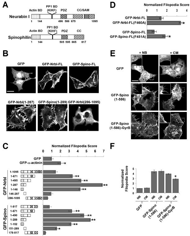
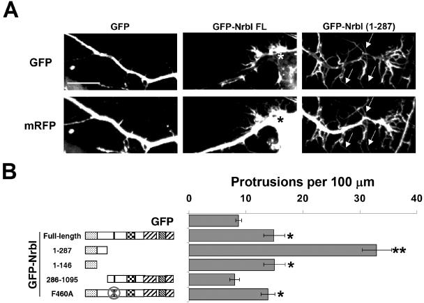
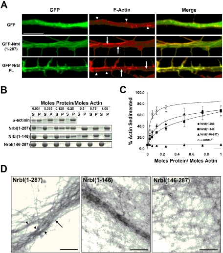
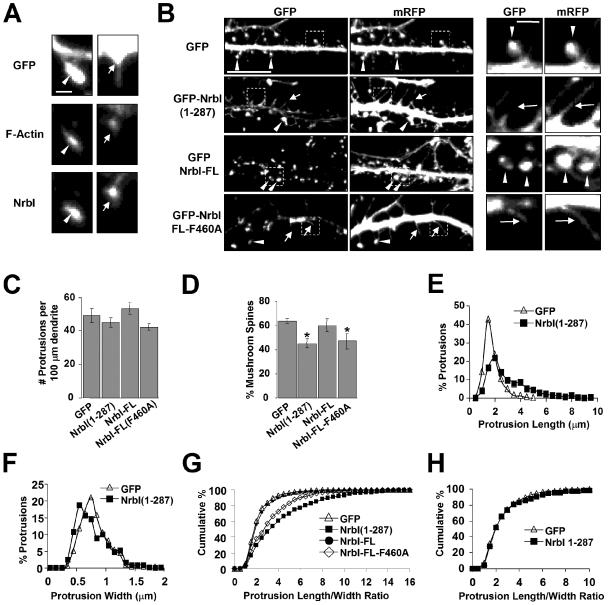
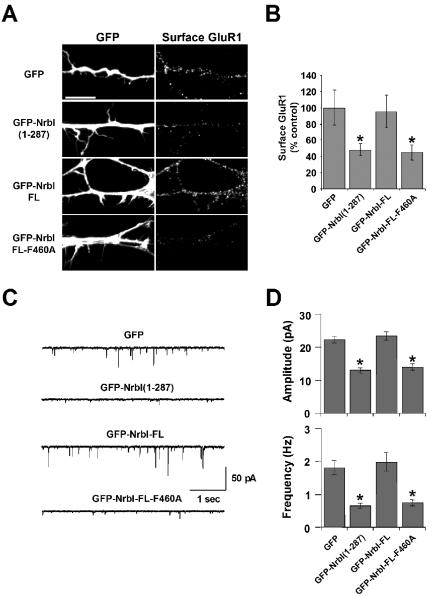
The majority of excitatory synapses in the mammalian brain form on filopodia and spines, actin-rich membrane protrusions present on neuronal dendrites. The biochemical events that induce filopodia and remodel these structures into dendritic spines remain poorly understood. Here, we show that the neuronal actin- and protein phosphatase-1-binding protein, neurabin-I, promotes filopodia in neurons and nonneuronal cells. Neurabin-I actin-binding domain bundled F-actin, promoted filopodia, and delayed the maturation of dendritic spines in cultured hippocampal neurons. In contrast, dimerization of neurabin-I via C-terminal coiled-coil domains and association of protein phosphatase-1 (PP1) with neurabin-I through a canonical KIXF motif inhibited filopodia. Furthermore, the expression of a neurabin-I polypeptide unable to bind PP1 delayed the maturation of neuronal filopodia into spines, reduced the synaptic targeting of AMPA-type glutamate (GluR1) receptors, and decreased AMPA receptor-mediated synaptic transmission. Reduction of endogenous neurabin levels by interference RNA (RNAi)-mediated knockdown also inhibited the surface expression of GluR1 receptors. Together, our studies suggested that disrupting the functions of a cytoskeletal neurabin/PP1 complex enhanced filopodia and impaired surface GluR1 expression in hippocampal neurons, thereby hindering the morphological and functional maturation of dendritic spines.


Figures

Figure 6.
Figure 6.
Endogenous neurabin-I regulates AMPAR surface expression. (A) pNrbI-OFF was constructed by insertion of the NrbI shRNA hairpin loop in the pZOFF vector. In this vector shRNA expression is driven by an H1 PolIII promoter and GFP is separately expressed under the control of a CMV promoter. (B) Left panels, GFP-NrbI-FL or GFP-Spino-FL was expressed into COS7 cells, which were transfected with pZOFF vector alone or pNrbI-OFF, and protein levels were analyzed using anti-GFP Western blots. Right panels, equal protein loading was verified by immunoblotting with an anti-tubulin antibody. (C) On DIV15, hippocampal neurons were transfected with pNrbI-OFF, and on DIV26, cells were immunostained with anti-NrbI antibody and imaged for both GFP and NrbI staining. Scale bar, 10 μm. (D) Quantification of NrbI expression in cultured hippocampal neurons transfected on DIV14 and immunostained on DIV19. Data are presented as a ratio of the anti-NrbI fluorescence intensity in GFP-positive cells relative to pZOFF containing control cells ± SEM (n >10 for each condition; **p < 0.01 using the Student's t test). (E) Hippocampal neurons were transfected on DIV14 with pZOFF and pNrbI-OFF. On DIV19, surface GluR1 AMPA receptor (AMPAR) subunits were labeled on live cells as described in Materials and Methods. Cells were imaged for GFP and GluR1 staining. (F) Quantification of surface GluR1 levels is shown as a fraction of the surface GluR1 fluorescence intensity in GFP-positive cells relative to pZOFF containing control cells ± SEM (n >30 for each condition; *p < 0.05 using the Student's t test). Scale bar, 10 μm.
Figure 1.
Figure 1.
Multiple protein-interaction domains in neurabin-I and spinophilin regulate the morphology of COS7 cells. (A) The schematic representation of neurabin-I (NrbI) and spinophilin/neurabin-II (Spino) highlights the actin-binding domain (Actin BD), PP1-binding motifs (PP1 BD; KIKF in NrbI and KIHF in Spino), PDZ domain, and the coiled-coil (CC) and sterile alpha motif (SAM) domains, which mediate oligomerization. The numbers indicate amino acids that flank these protein interaction domains. (B) COS7 cells were transfected with GFP-NrbI and GFP-Spino plasmids, fixed after 16 h, and imaged. Scale bar, 20 μm. (C) Filopodia induced by GFP-neurabins were quantified and normalized to control GFP-expressing cells. Results are shown ± SEM; *p < 0.05 and **p < 0.005 using Student's t test. (D) Filopodia induced by NrbI and Spino proteins unable to bind PP1 because of the F-to-A substitutions within the PP1-binding motif were compared with control GFP-expressing cells as described above. *p < 0.05 using Student's t test. (E) COS7 cells expressing GFP-Spino(1–586) or GFP-Spino(1–586)-GyrB were incubated with either novobiocin (NB) or coumermycin (CM) for 16 h before imaging. Scale bar, 20 μm. Inset represent higher magnification of regions of these cells marked by a dashed box. Scale bar, 5 μm. (F) Data from the experiment shown in E were quantified as described for C. *p < 0.05 using Student's t test.
Figure 2.
Figure 2.
Neurabins induce dendritic filopodia in hippocampal neurons. (A) Cultured hippocampal neurons (DIV7) were cotransfected with plasmids encoding mRFP along with GFP, GFP-NrbI-FL, or GFP-NrbI(1–287). Representative dendrites of DIV8 neurons cotransfected with the GFP fusion protein (top panels) and mRFP (bottom panels) are shown. Arrows indicate filopodia. Asterisk (*) indicates a filopodial “burst.” Scale bar, 10 μm. (B) Quantification of dendritic protrusions expressed per length of dendrite is shown for experiments conducted as described in A. The data for the number of protrusions per 100 μm of dendrite are shown ± SEM; *p < 0.05, **p < 0.005 compared with GFP controls, Student's t test.
Figure 3.
Figure 3.
The N-terminal domain of neurabin-I reorganizes the neuronal actin cytoskeleton in vivo and bundles actin in vitro. (A) Cultured hippocampal neurons (DIV8) expressing GFP-neurabins were fixed and F-actin was stained with Alexa-568-phalloidin. Representative GFP fluorescence, phalloidin staining, and merged images are shown. Arrowheads highlight F-actin puncta and arrows indicate linear bundles of actin filaments. Scale bar, 5 μm. (B) Purified F-actin was mixed with α-actinin or His-NrbI proteins at the increasing molar ratios and low-speed centrifugation separated F-actin filaments in the supernatant (S) from bundled F-actin in the pellet (P). Supernatants and pellets were subjected to SDS-PAGE, and the gels were stained with Coomassie blue. Coomassie-stained actin is shown. (C) Quantification of results in B is shown as the percent of total F-actin sedimented at various His-NrbI/actin molar ratios (±SEM; n = 4). (D) The bundles of F-actin filaments induced by His-NrbI proteins were examined by electron microscopy. Arrow points to a “loose” F-actin bundle consisting of several smaller bundles (arrowheads). Scale bars, 200 nm.
Figure 4.
Figure 4.
Neurabin-I regulates spine development in hippocampal neurons. (A) Hippocampal neurons (DIV35) expressing GFP were stained with Alexa568-phalloidin and immunostained for endogenous NrbI using an anti-NrbI antibody. An actin-rich NrbI-containing spine (left) and a dendritic filopodia with NrbI localized at the base (right) are shown. Scale bar, 1 μm. (B) Hippocampal neurons (DIV12) were cotransfected with plasmids that expressed mRFP and GFP-NrbI fusion proteins, fixed at DIV26, and imaged. Arrowheads indicate spines and arrows point to filopodia. Scale bar, 10 μm. Right panels show the enlarged images of the areas indicated by the dotted squares. Scale bar, 1 μm. (C) Using the mRFP channel, the number of protrusions per unit length of dendrite was measured following the expression of the indicated GFP-NrbI proteins. (D) Dendritic protrusions of neurons transfected with indicated NrbI proteins were defined as either mushroom-shaped spines or nonspines/filopodia (see Materials and Methods for details) and expressed as the percentage of protrusions that are spines. *p < 0.05, Student's t test. (E) Length of individual protrusions was measured and the results were presented as histograms. (F) Histogram of protrusion width. (G) The length/width ratios of dendritic protrusions expressing the indicated GFP-fusion proteins were calculated, and results are presented as cumulative frequency distributions. (H) Hippocampal neurons were cotransfected with mRFP and GFP-NrbI(1–287) at DIV28 and fixed at DIV30. Results from pooled experiments were quantified, and length/width ratios are expressed as cumulative frequency distributions.
Figure 5.
Figure 5.
Neurabin-I regulates surface AMPA Receptor expression and excitatory synaptic transmission. (A) Hippocampal neurons were transfected on DIV14 with plasmids encoding the indicated GFP-NrbI proteins. On DIV17, surface GluR1 AMPA receptor (AMPAR) subunits were labeled on live cells by incubating with anti-GluR1 antibody directed against an extracellular epitope before fixation and immunostaining with fluorophore-conjugated secondary antibody. Cells were then imaged for GFP and surface GluR1 staining. Scale bar, 10 μm. (B) Quantification of surface GluR1 levels of pooled experiments shown in A with ± SEM (n = 12–15 for each transfection; *p < 0.05, Student's t test). (C) Representative traces of mEPSCs recorded from DIV16 hippocampal neurons expressing GFP (top) or the indicated GFP-NrbI proteins are shown. (D) mEPSC amplitudes (top) and frequencies (bottom) are shown with SEM determined using Student's t test (n = 8–11 for each construct; *p < 0.05).

Similar articles

Cited by

References

    1. Allen, P. B., Greenfield, A. T., Svenningsson, P., Haspeslagh, D. C., and Greengard, P. (2004). Phactrs 1–4, a family of protein phosphatase 1 and actin regulatory proteins. Proc. Natl. Acad. Sci. USA 101, 7187–7192. - PMC - PubMed
    1. Allen, P. B., Ouimet, C. C., and Greengard, P. (1997). Spinophilin, a novel protein phosphatase 1 binding protein localized to dendritic spines. Proc. Natl. Acad. Sci. USA 94, 9956–9961. - PMC - PubMed
    1. Barnes, A. P., Smith, F. D., 3rd, VanDongen, H. M., VanDongen, A. M., and Milgram, S. L. (2004). The identification of a second actin-binding region in spinophilin/neurabin II. Brain Res. Mol. Brain Res. 124, 105–113. - PubMed
    1. Beattie, E. C., Carroll, R. C., Yu, X., Morishita, W., Yasuda, H., von Zastrow, M., and Malenka, R. C. (2000). Regulation of AMPA receptor endocytosis by a signaling mechanism shared with LTD. Nat. Neurosci. 3, 1291–1300. - PubMed
    1. Bonhoeffer, T., and Yuste, R. (2002). Spine motility. Phenomenology, mechanisms, and function. Neuron 35, 1019–1027. - PubMed

Publication types

MeSH terms

LinkOut - more resources