. 2005 May;16(5):2363-71.
doi: 10.1091/mbc.e04-10-0878. Epub 2005 Mar 2.

FXYD3 (Mat-8), a new regulator of Na,K-ATPase

Affiliations

FXYD3 (Mat-8), a new regulator of Na,K-ATPase

Gilles Crambert et al. Mol Biol Cell. 2005 May.

Abstract

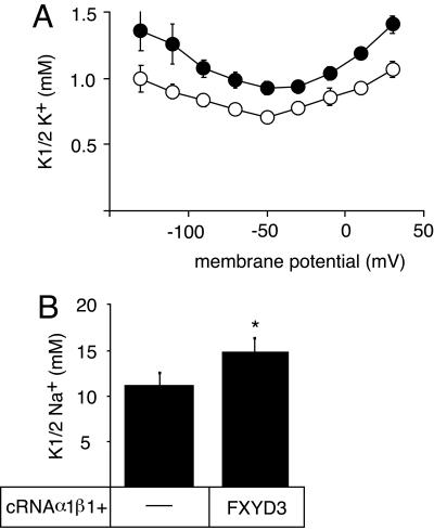
Four of the seven members of the FXYD protein family have been identified as specific regulators of Na,K-ATPase. In this study, we show that FXYD3, also known as Mat-8, is able to associate with and to modify the transport properties of Na,K-ATPase. In addition to this shared function, FXYD3 displays some uncommon characteristics. First, in contrast to other FXYD proteins, which were shown to be type I membrane proteins, FXYD3 may have a second transmembrane-like domain because of the presence of a noncleavable signal peptide. Second, FXYD3 can associate with Na,K- as well as H,K-ATPases when expressed in Xenopus oocytes. However, in situ (stomach), FXYD3 is associated only with Na,K-ATPase because its expression is restricted to mucous cells in which H,K-ATPase is absent. Coexpressed in Xenopus oocytes, FXYD3 modulates the glycosylation processing of the beta subunit of X,K-ATPase dependent on the presence of the signal peptide. Finally, FXYD3 decreases both the apparent affinity for Na+ and K+ of Na,K-ATPase.


Figures

Figure 1.
Figure 1.
Biosynthesis of FXYD3. (A) Sequence comparison between mouse and rat FXYD3 and FXYD4. The vertical bar indicates the putative cleavage site of the signal peptide. The FXYD motif is indicated. The transmembrane domain is boxed. (B) Wild-type FXYD3 and the Nt-Δ22 cRNAs were translated in vitro in the absence (lanes 1 and 3) or presence (lanes 2 and 4) of canine pancreatic microsomes and [35S]methionine. Mouse FXYD3 cRNA (2 ng) was injected into Xenopus oocytes and pulse labeled with [35S]methionine for 24 h (lane 5) followed by a 48-h chase period (lane 6). In vitro-translated samples or oocyte microsomes were subjected to SDS-tricine gel electrophoresis and revealed by fluorography. Proteins from microsomes of FXYD3-expressing oocytes (50 μg) (lane 7) or from mouse stomach microsomes (50 μg) (lane 8) or from nontransfected or FXYD3-transfected HEK cells (50 μg) (lanes 9 and 10) were resolved on SDS-tricine gels and subjected to Western blot analysis by using an anti-FXYD4 antibody (1/250). Indicated is the molecular mass of FXYD3 and the Nt-Δ22 mutant. (C) Mouse FXYD3, FXYD4, or FXYD4/FXYD3 cRNAs (2 ng) were injected into Xenopus oocytes and pulse labeled with [35S]methionine for 6 h followed by a 24- and a 48-h chase period. Oocyte microsomes were subjected to SDS-tricine gel electrophoresis and revealed by fluorography. (D) Mouse FXYD3 and V5N mutant cRNAs were translated in vitro in the absence (lanes 1 and 3) or presence (lanes 2 and 4) of canine pancreatic microsomes. In vitro-translated samples were subjected to SDS-tricine gel electrophoresis and revealed by fluorography. One of two similar experiments is shown.
Figure 2.
Figure 2.
Association of FXYD3 with P-type ATPases and other transporters in Xenopus oocytes, and effect on the glycosylation processing. (A) Oocytes were injected with rat Na,K-ATPase (NKA) cRNA (10 ng of α1 and 1 ng of β1 cRNAs, lanes 1–6) or rat colonic H,K-ATPase (colonic HKA) cRNA (10 ng of α1 and 1 ng of Bufo bladder β cRNAs, lanes 7–12), or human SERCA2a cRNA (5 ng, lanes 13–16) in the absence or presence of 2 ng of FXYD3 cRNA. After cRNA injection, oocytes were labeled with [35S]methionine for 6 h followed by a 24- and 48-h chase period, and digitonin extracts were prepared. Nondenaturing immunoprecipitation with antibodies against the Na,K-ATPase α subunit, the colonic H,K-ATPase, or SERCA2a was performed, and the immunoprecipitates were resolved by SDS-PAGE and revealed by fluorography. (B) Oocytes were injected with rat Na,K-ATPase (NKA) cRNA (10 ng of α1 and 1ngof β1 cRNAs, lanes 1–3 and 7–9) or rabbit, gastric H,K-ATPase cRNA (HKA) (10 ng of α1 and 1 ng of β1 cRNAs, lanes 4–6 and 10–12) in the presence of 2 ng of FXYD4 (lanes 1–6) or FXYD3 (lanes 7–12). Nondenaturing immunoprecipitations were performed with antibodies against the Na,K-ATPase α subunit or the gastric H,K-ATPase. (C) Oocytes were injected with 5 ng of Glu-2 transporter cRNA (lanes 1–6) or 5 ng of FLAG-Aquaporin-2 cRNA (lanes 7–12) in the absence (lanes 1–3 and 7–9) or presence of FXYD3 (lanes 4–6 and 10–12). Digitonin extracts were prepared after the pulse and the chase periods and were immunoprecipitated under nondenaturing conditions with a Glut-2 antibody (lanes 1–6) or a FLAG antibody (lanes 7–12). cg, core glycosylated form; fg, fully glycosylated form. One of 2–20 experiments is shown.
Figure 3.
Figure 3.
Effect of FXYD3 on the glycosylation processing and cell surface expression of the Na,K-ATPase in Xenopus oocytes. (A and B) Rat Na,K-ATPase α1 (10 ng) and Xenopus FLAGβ1 (2 ng) cRNAs (A) or rat α1 and β1 (B) cRNAs were injected into oocytes in the absence or presence of mouse FXYD3 (2 ng) or Nt-Δ22 mutant (4 ng) cRNAs, and pulse labeled for 6 h followed by various chase periods. Microsomes were prepared and subjected to nondenaturating immunoprecipitations by using an antibody against Na,K-ATPase α subunit. Immunoprecipitates were resolved on 5–13% SDS-PAG and revealed by fluorography. (C) Three days after injection as described in A, intact noninjected (ni) or cRNA-injected oocytes were subjected to a radioimmunolabeling assay by using a 125I-labeled anti-FLAG antibody. Shown are means ± SE from seven to nine oocytes. (D) Three days after injection as described in B, the K+-activated Na,K-pump current was measured on intact oocytes by the two-electrode voltage-clamp technique as described in Materials and Methods. Shown are means ± SE of five to six oocytes from two batches of oocytes.
Figure 4.
Figure 4.
FXYD3 is associated with Na,K-ATPase in stomach epithelial cell. (A) Aliquots (40 μg of protein) from stomach epithelial microsomes were directly loaded on a tricine-gel (lane 1) or first immunoprecipitated (400 μg of protein) with an antibody against Na,K-ATPase α subunit (lane 2) or against gastric H,K-ATPase α subunit (6H) (lane 3) under nondenaturating conditions. Proteins were transferred onto nitrocellulose, and Western blot analysis was performed using an anti-FXYD4 antibody. (B and C) Mouse stomach slices were used for simple or double immunostaining with an anti FXYD3 antibody (Bc, Cb, and Ce, green) and with either a monoclonal anti-H,K-ATPase antibody (HK9 in Ba) or monoclonal anti-Na,K-ATPase antibodies (6H, in Bb; 9-A5, in Ca and Cd, red). After merging (Cc and Cf), colocalization is indicated in yellow. Magnification, 40× (Ba, Bb, Bd, Ca, Cb, and Cc) and 63× (Cd, Ce, and Cf).
Figure 5.
Figure 5.
Effects of FXYD3 on the transport and enzymatic properties of Na,K-ATPase. (A) Three days after injection of rat Na,K-ATPase α1 (10 ng) and β1 (1 ng) cRNAs in the absence or presence of FXYD3 (2 ng) cRNA, K+-activation constants (K½ K+) of the Na,K-ATPase were determined in the presence of 90 mM external Na+. Open circles, Na,K-ATPase alone; closed circles, Na,K-ATPase plus FXYD3. Shown are means ± SE of 10 oocytes from two different batches. Phosphate-buffered saline <0.05 at all membrane potentials. (B) Two days after injection of rat α1 (10 ng) and β1 (1 ng) cRNAs in the absence or presence of FXYD3 (2 ng) cRNA, the epithelial Na+ channel subunit cRNAs (α, β, and γ, 0.3 ng each) were injected. After overnight incubation, Na+ activation constants (K½ Na+) were determined at–50 mV. Shown are means ± SE from 11 oocytes from three different batches. *p < 0.01. For electrophysiological measurements, endogenous, oocyte Na,K-ATPase was inhibited by the presence of 1 μM ouabain.

Similar articles

Cited by

References

    1. Arystarkhova, E., Wetzel, R. K., Asinovski, N. K., and Sweadner, K. J. (1999). The γ subunit modulates Na+ and K+ affinity of the renal Na,K-ATPase. J. Biol. Chem. 274, 33183–33185. - PubMed
    1. Béguin, P., Crambert, G., Guennoun, S., Garty, H., Horisberger, J.-D., and Geering, K. (2001). CHIF, a member of the FXYD protein family, is a regulator of Na,K-ATPase distinct from the γ-subunit. EMBO J. 20, 3993–4002. - PMC - PubMed
    1. Béguin, P., Crambert, G., Monnet-Tschudi, F., Uldry, M., Horisberger, J.-D., Garty, H., and Geering, K. (2002). FXYD7 is a brain-specific regulator of Na,K-ATPase α1-β isozymes. EMBO J. 21, 3264–3273. - PMC - PubMed
    1. Béguin, P., Wang, X. Y., Firsov, D., Puoti, A., Claeys, D., Horisberger, J. D., and Geering, K. (1997). The γ subunit is a specific component of the Na,K-ATPase and modulates its transport properties. EMBO J. 16, 4250–4260. - PMC - PubMed
    1. Bendtsen, J. D., Nielsen, H., von Heijne, G., and Brunak, S. (2004). Improved prediction of signal peptides: SignalP 3.0. J. Mol. Biol. 340, 783–795. - PubMed

Publication types

MeSH terms