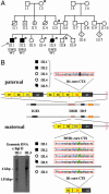
Microdeletion of target sites for insulator protein CTCF in a chromosome 11p15 imprinting center in Beckwith-Wiedemann syndrome and Wilms' tumor
- PMID: 15743916
- PMCID: PMC554791
- DOI: 10.1073/pnas.0500037102
Microdeletion of target sites for insulator protein CTCF in a chromosome 11p15 imprinting center in Beckwith-Wiedemann syndrome and Wilms' tumor
Abstract
We have analyzed several cases of Beckwith-Wiedemann syndrome (BWS) with Wilms' tumor in a familial setting, which give insight into the complex controls of imprinting and gene expression in the chromosome 11p15 region. We describe a 2.2-kbp microdeletion in the H19/insulin-like growth factor 2 (IGF2)-imprinting center eliminating three target sites of the chromatin insulator protein CTCF that we believe here is necessary, but not sufficient, to cause BWS and Wilms' tumor. Maternal inheritance of the deletion is associated with IGF2 loss of imprinting and up-regulation of IGF2 mRNA. However, in at least one affected family member a second genetic lesion (a duplication of maternal 11p15) was identified and accompanied by a further increase in IGF2 mRNA levels 35-fold higher than control values. Our results suggest that the combined effects of the H19/IGF2-imprinting center microdeletion and 11p15 chromosome duplication were necessary for manifestation of BWS.
Figures


Similar articles
-
Microdeletions in the human H19 DMR result in loss of IGF2 imprinting and Beckwith-Wiedemann syndrome.Nat Genet. 2004 Sep;36(9):958-60. doi: 10.1038/ng1410. Epub 2004 Aug 15. Nat Genet. 2004. PMID: 15314640
-
Different mechanisms cause imprinting defects at the IGF2/H19 locus in Beckwith-Wiedemann syndrome and Wilms' tumour.Hum Mol Genet. 2008 May 15;17(10):1427-35. doi: 10.1093/hmg/ddn031. Epub 2008 Feb 1. Hum Mol Genet. 2008. PMID: 18245780
-
Analysis of the IGF2/H19 imprinting control region uncovers new genetic defects, including mutations of OCT-binding sequences, in patients with 11p15 fetal growth disorders.Hum Mol Genet. 2010 Mar 1;19(5):803-14. doi: 10.1093/hmg/ddp549. Epub 2009 Dec 9. Hum Mol Genet. 2010. PMID: 20007505
-
Inherited and Sporadic Epimutations at the IGF2-H19 locus in Beckwith-Wiedemann syndrome and Wilms' tumor.Endocr Dev. 2009;14:1-9. doi: 10.1159/000207461. Epub 2009 Feb 27. Endocr Dev. 2009. PMID: 19293570 Review.
-
Role of genomic imprinting in Wilms' tumour and overgrowth disorders.Med Pediatr Oncol. 1996 Nov;27(5):470-5. doi: 10.1002/(SICI)1096-911X(199611)27:5<470::AID-MPO14>3.0.CO;2-E. Med Pediatr Oncol. 1996. PMID: 8827076 Review.
Cited by
-
De novo mutations in the genome organizer CTCF cause intellectual disability.Am J Hum Genet. 2013 Jul 11;93(1):124-31. doi: 10.1016/j.ajhg.2013.05.007. Epub 2013 Jun 6. Am J Hum Genet. 2013. PMID: 23746550 Free PMC article.
-
CTCFBSDB: a CTCF-binding site database for characterization of vertebrate genomic insulators.Nucleic Acids Res. 2008 Jan;36(Database issue):D83-7. doi: 10.1093/nar/gkm875. Epub 2007 Nov 2. Nucleic Acids Res. 2008. PMID: 17981843 Free PMC article.
-
Epigenetics and neural stem cell commitment.Neurosci Bull. 2007 Jul;23(4):241-8. doi: 10.1007/s12264-007-0036-8. Neurosci Bull. 2007. PMID: 17687400 Free PMC article. Review.
-
Functional roles of CTCF in breast cancer.BMB Rep. 2017 Sep;50(9):445-453. doi: 10.5483/bmbrep.2017.50.9.108. BMB Rep. 2017. PMID: 28648147 Free PMC article.
-
Mutation of a barrier insulator in the human ankyrin-1 gene is associated with hereditary spherocytosis.J Clin Invest. 2010 Dec;120(12):4453-65. doi: 10.1172/JCI42240. Epub 2010 Nov 22. J Clin Invest. 2010. PMID: 21099109 Free PMC article.
References
-
- Weksberg, R., Smith, A. C., Squire, J. & Sadowski, P. (2003) Hum. Mol. Genet. 12, Spec. no. 1, R61–R68. - PubMed
-
- Rainier, S., Johnson, L. A., Dobry, C. J., Ping, A. J., Grundy, P. E. & Feinberg, A. P. (1993) Nature 362, 747–749. - PubMed
-
- Soejima, H., Nakagawachi, T., Zhao, W., Higashimoto, K., Urano, T., Matsukura, S., Kitajima, Y., Takeuchi, M., Nakayama, M., Oshimura, M., et al. (2004) Oncogene 23, 4380–4388. - PubMed
-
- Banet, G., Bibi, O., Matouk, I., Ayesh, S., Laster, M., Kimber, K. M., Tykocinski, M., de Groot, N., Hochberg, A. & Ohana, P. (2000) Mol. Biol. Rep. 27, 157–165. - PubMed
Publication types
MeSH terms
Substances
LinkOut - more resources
Full Text Sources
Other Literature Sources
Medical
Miscellaneous

