. 2005 Apr 5;102(14):5162-7.
doi: 10.1073/pnas.0408536102. Epub 2005 Mar 2.

Borrelia burgdorferi sigma54 is required for mammalian infection and vector transmission but not for tick colonization

Affiliations

Borrelia burgdorferi sigma54 is required for mammalian infection and vector transmission but not for tick colonization

Mark A Fisher et al. Proc Natl Acad Sci U S A. .

Abstract

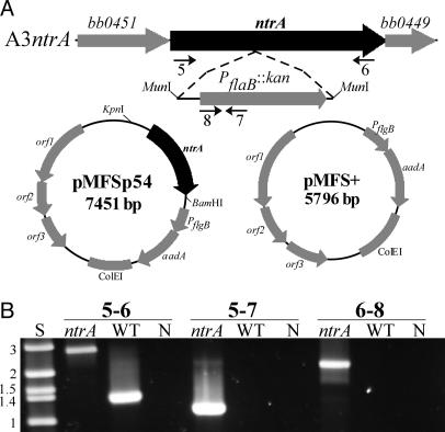
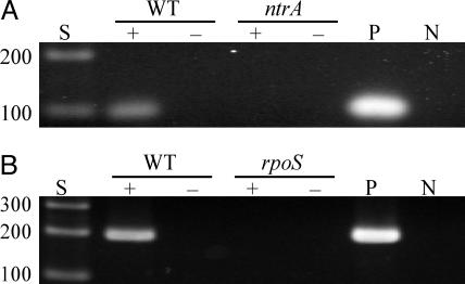
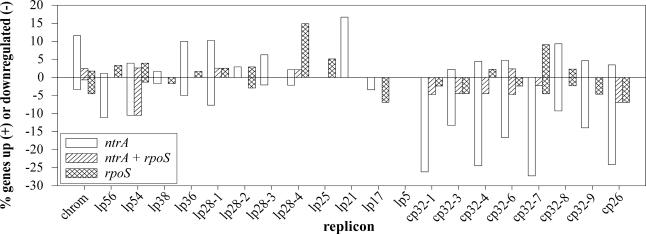
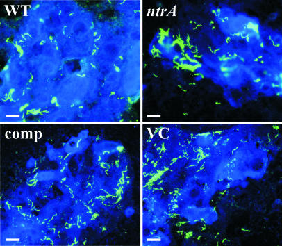
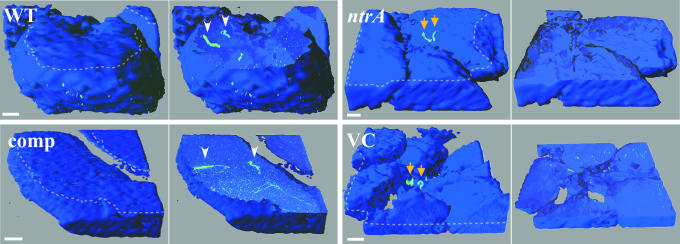
Previous studies have shown that a sigma54-sigma(S) cascade regulates the expression of a few key lipoproteins in Borrelia burgdorferi, the agent of Lyme disease. Here, we demonstrate that these sigma factors, both together and independently, regulate a much more extensive number of genes and cellular processes. Microarray analyses of sigma54 and sigma(S) mutant strains identified 305 genes regulated by sigma54 and 145 regulated by sigma(S), whereas the sigma54-sigma(S) regulatory cascade appears to control 48 genes in B. burgdorferi. In silico analyses revealed that nearly 80% of genes with altered expression in the sigma54 mutant were linked to potential sigma54-dependent promoters. Many sigma54-regulated genes are expressed in vivo, and through genetic complementation of the mutant, we demonstrated that sigma54 was required by B. burgdorferi to infect mammals. Surprisingly, sigma54 mutants were able to infect Ixodes scapularis ticks and be maintained for at least 24 wk after infection, suggesting the sigma54-sigma(S) regulatory network was not involved in long-term survival in ticks. However, sigma54 mutants did not enter the salivary glands during tick feeding, indicating that sigma54-regulated genes were involved in the transmission process.


Figures

Fig. 1.
Fig. 1.
Disruption of ntrA and complementation of the σ54 mutant. (A) Flanking ORFs and insertion point of the PflaB::kan cassette in strain A3ntrA are shown above the complementation (pMFSp54) and VC (pMFS+) plasmids. Numbered arrows indicate the relative positions of primers used for PCR confirmation of the mutant. (B) PCR analyses to confirm ntrA disruption in A3ntrA. Size standards (S) in kbp and negative controls (N) are indicated.
Fig. 2.
Fig. 2.
RT-PCR analysis of ntrA and rpoS expression. RNA isolated from each strain was converted to cDNA and PCR-amplified with primers specific for ntrA or rpoS. (A) Expression of ntrA in B31-A3 (WT) and A3ntrA (ntrA, σ54 mutant). (B) Expression of rpoS in B31-A3 (WT) and A3rpoS (rpoS, σS mutant). Size standards (S) are in bp, and positive (P, B31-A3 DNA) and negative (N) controls are indicated. The presence or absence of reverse transcriptase is indicated by + or –.
Fig. 3.
Fig. 3.
Distribution of regulated genes by replicon. Microarray data were categorized by replicon and sigma factor regulatory group and are shown as percent of ORFs significantly changed on each replicon. ntrA, genes regulated only by σ54; ntrA + rpoS, genes regulated by both σ54 and σS; rpoS, genes regulated only by σS.
Fig. 4.
Fig. 4.
IFA of midguts from fed ticks 24 wk after infection. Midguts isolated from ticks infected with A3-Gm (WT), A3ntrA-Gm (ntrA, σ54 mutant), ntrA-comp, and ntrA-VC strains were examined by IFA. Spirochetes (green) were detected by fluorescent microscopy with rabbit anti-B. burgdorferi primary and Alexa Fluor 488-labeled anti-rabbit secondary antisera. (Scale bars, 20 μm.)
Fig. 5.
Fig. 5.
3D surface and cutaway projections of salivary gland ascini. Salivary glands were isolated from partially fed ticks infected with A3-Gm (WT), A3ntrA-Gm (ntrA, σ54 mutant), ntrA-comp, and ntrA-VC strains. The left image in each case shows the outer surface of intact ascini, and the right image shows a computer-generated internal section of the corresponding structure. The cutting plane used in the right image is denoted by the dotted line in the left image. Arrowheads indicate spirochetes (green) located within ascini, and arrows denote those on the exterior of the structure. Spirochetes were detected by confocal microscopy using rabbit anti-B. burgdorferi primary and Alexa Fluor 488-labeled anti-rabbit secondary antisera, and salivary glands were stained with DRAQ5. (Scale bars, 10 μm.)

Comment in

Similar articles

Cited by

References

    1. Fraser, C. M., Casjens, S., Huang, W. M., Sutton, G. G., Clayton, R., Lathigra, R., White, O., Ketchum, K. A., Dodson, R., Hickey, E. K., et al. (1997) Nature 390, 580–586. - PubMed
    1. Casjens, S., Palmer, N., van Vugt, R., Huwang, W. M., Stevenson, B., Rosa, P., Lathigra, R., Sutton, G. G., Peterson, J., Dodson, R., et al. (2000) Mol. Microbiol. 35, 490–516. - PubMed
    1. Narasimhan, S., Camaino, M. J., Liang, F. T., Santiago, F., Laskowski, M., Philipp, M. T., Pachner, A. R., Radolf, J. D. & Fikrig, E. (2003) Proc. Natl. Acad. Sci. USA 100, 15953–15958. - PMC - PubMed
    1. Narasimhan, S., Santiago, F., Koski, R. A., Brei, B., Anderson, J. F., Fish, D. & Fikrig, E. (2002) J. Bacteriol. 184, 3122–3125. - PMC - PubMed
    1. Brooks, C. S., Hefty, P. S., Jolliff, S. E. & Akins, D. R. (2003) Infect. Immun. 71, 3371–3383. - PMC - PubMed

MeSH terms

LinkOut - more resources