

Crystal structure of low-molecular-weight protein tyrosine phosphatase from Mycobacterium tuberculosis at 1.9-A resolution
- PMID: 15743966
- PMCID: PMC1064030
- DOI: 10.1128/JB.187.6.2175-2181.2005
Crystal structure of low-molecular-weight protein tyrosine phosphatase from Mycobacterium tuberculosis at 1.9-A resolution
Abstract
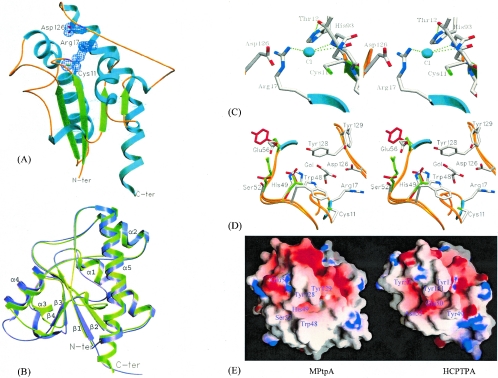
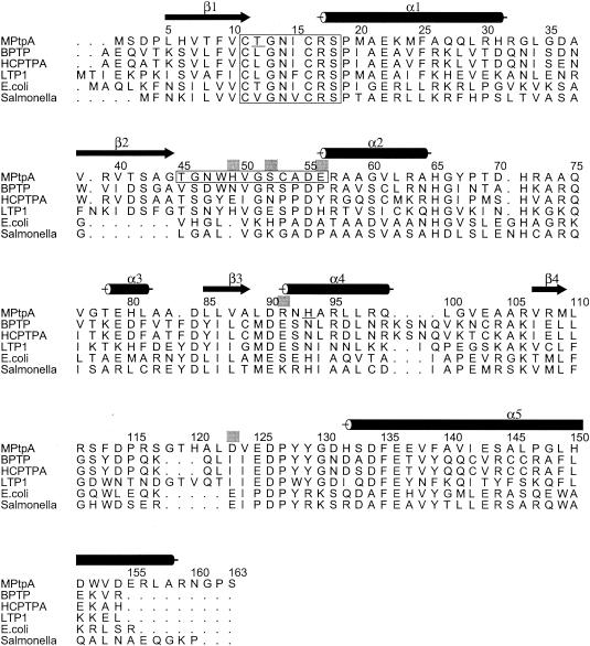
The low-molecular-weight protein tyrosine phosphatase (LMWPTPase) belongs to a distinctive class of phosphotyrosine phosphatases widely distributed among prokaryotes and eukaryotes. We report here the crystal structure of LMWPTPase of microbial origin, the first of its kind from Mycobacterium tuberculosis. The structure was determined to be two crystal forms at 1.9- and 2.5-A resolutions. These structural forms are compared with those of the LMWPTPases of eukaryotes. Though the overall structure resembles that of the eukaryotic LMWPTPases, there are significant changes around the active site and the protein tyrosine phosphatase (PTP) loop. The variable loop forming the wall of the crevice leading to the active site is conformationally unchanged from that of mammalian LMWPTPase; however, differences are observed in the residues involved, suggesting that they have a role in influencing different substrate specificities. The single amino acid substitution (Leu12Thr [underlined below]) in the consensus sequence of the PTP loop, CTGNICRS, has a major role in the stabilization of the PTP loop, unlike what occurs in mammalian LMWPTPases. A chloride ion and a glycerol molecule were modeled in the active site where the chloride ion interacts in a manner similar to that of phosphate with the main chain nitrogens of the PTP loop. This structural study, in addition to identifying specific mycobacterial features, may also form the basis for exploring the mechanism of the substrate specificities of bacterial LMWPTPases.
Figures
Similar articles
-
Mycobacterium tuberculosis protein tyrosine phosphatase PtpB structure reveals a diverged fold and a buried active site.Structure. 2005 Nov;13(11):1625-34. doi: 10.1016/j.str.2005.07.017. Structure. 2005. PMID: 16271885
-
Solution structure of a low-molecular-weight protein tyrosine phosphatase from Bacillus subtilis.J Bacteriol. 2006 Feb;188(4):1509-17. doi: 10.1128/JB.188.4.1509-1517.2006. J Bacteriol. 2006. PMID: 16452434 Free PMC article.
-
Crystal structure of a human low molecular weight phosphotyrosyl phosphatase. Implications for substrate specificity.J Biol Chem. 1998 Aug 21;273(34):21714-20. doi: 10.1074/jbc.273.34.21714. J Biol Chem. 1998. PMID: 9705307
-
Structure and catalytic properties of protein tyrosine phosphatases.Ann N Y Acad Sci. 1995 Sep 7;766:18-22. doi: 10.1111/j.1749-6632.1995.tb26644.x. Ann N Y Acad Sci. 1995. PMID: 7486654 Review. No abstract available.
-
Crystal structure of the Yersinia tyrosine phosphatase.Trends Microbiol. 1995 Apr;3(4):125-7. doi: 10.1016/s0966-842x(00)88898-8. Trends Microbiol. 1995. PMID: 7613751 Free PMC article. Review. No abstract available.
Cited by
-
Voltage sensitive phosphatases: emerging kinship to protein tyrosine phosphatases from structure-function research.Front Pharmacol. 2015 Jan 10;6:20. doi: 10.3389/fphar.2015.00020. eCollection 2015. Front Pharmacol. 2015. PMID: 25713537 Free PMC article. Review.
-
Effects of SDS on the activity and conformation of protein tyrosine phosphatase from thermus thermophilus HB27.Sci Rep. 2020 Feb 21;10(1):3195. doi: 10.1038/s41598-020-60263-4. Sci Rep. 2020. PMID: 32081966 Free PMC article.
-
Wzb of Vibrio vulnificus represents a new group of low-molecular-weight protein tyrosine phosphatases with a unique insertion in the W-loop.J Biol Chem. 2021 Jan-Jun;296:100280. doi: 10.1016/j.jbc.2021.100280. Epub 2021 Jan 12. J Biol Chem. 2021. PMID: 33450227 Free PMC article.
-
Crystal structure and putative substrate identification for the Entamoeba histolytica low molecular weight tyrosine phosphatase.Mol Biochem Parasitol. 2014 Jan;193(1):33-44. doi: 10.1016/j.molbiopara.2014.01.003. Epub 2014 Feb 15. Mol Biochem Parasitol. 2014. PMID: 24548880 Free PMC article.
-
New potential eukaryotic substrates of the mycobacterial protein tyrosine phosphatase PtpA: hints of a bacterial modulation of macrophage bioenergetics state.Sci Rep. 2015 Mar 6;5:8819. doi: 10.1038/srep08819. Sci Rep. 2015. PMID: 25743628 Free PMC article.
References
-
- Acta Crystallographica D. 1994. Collaborative computational project number 4. The CCP4 suite: programs for protein crystallography. Acta Crystallogr. D 50:760-763. - PubMed
-
- Barford, D., A. J. Flint, and N. K. Tonks. 1994. The crystal structure of human protein tyrosine phosphatase 1B. Science 263:1397-1404. - PubMed
-
- Barton, G. J. 1993. ALSCRIPT: a tool to format multiple sequence alignments. Protein Eng. 6:37-40. - PubMed
-
- Bilwes, A. M., J. Den Hertog, T. Hunter, and J. P. Noel. 1996. Structural basis for inhibition of receptor protein-tyrosine phosphatase-alpha by dimerization. Nature 382:555-559. - PubMed
Publication types
MeSH terms
Substances
Associated data
- Actions
- Actions
Grants and funding
LinkOut - more resources
Full Text Sources
