Review
. 2005 Mar;26(3):150-6.
doi: 10.1016/j.it.2004.11.014.

A stromal address code defined by fibroblasts

Affiliations
Review

A stromal address code defined by fibroblasts

Greg Parsonage et al. Trends Immunol. 2005 Mar.

Abstract

To navigate into and within tissues, leukocytes require guidance cues that enable them to recognize which tissues to enter and which to avoid. Such cues are partly provided at the time of extravasation from blood by an endothelial address code on the luminal surface of the vascular endothelium. Here, we review the evidence that fibroblasts help define an additional stromal address code that directs leukocyte behaviour within tissues. We examine how this stromal code regulates site-specific leukocyte accumulation, differentiation and survival in a variety of physiological stromal niches, and how the aberrant expression of components of this code in the wrong tissue at the wrong time contributes to the persistence of chronic inflammatory diseases.


Figures

Figure 1
Figure 1
Vascular codes provide for compartmentalization within the immune system. During the development of an immune response, immature naïve T cells (red dotted arrows) and B cells (green dotted arrows), which have been educated in the thymus and bone marrow, respectively, enter lymph nodes through the high endothelial venules (HEVs) using the endothelial entry code CCR7 and L-selectin (area codes are shown in grey). Antigen-experienced B cells (green arrows) leave the lymph node and migrate into specific niches, such as the red pulp of the spleen and the bone marrow, using the endothelial entry code CXCR4. Antigen-experienced central memory T cells continue to recirculate back to the lymph nodes via the efferent lymphatics and thoracic duct (red arrows; dot and dash). Effector memory T cells preferentially recirculate to the tissue in which they were initially activated; for example, if they are activated in a lymph node draining the skin they acquire the skin entry code CCR4, CCR10, CLA (grey) and if activated in a lymph node draining the gut they acquire the gut entry code CCR9, α4β7. At the end of immune surveillance within tissues, memory T cells can recirculate back to the lymph nodes via the afferent lymphatics (red arrows).
Figure 2
Figure 2
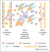
Leukocyte–stromal interactions are involved at every stage in lymphocyte recirculation. The molecular basis by which leukocytes leave the circulation and migrate across endothelium has been well studied (step 1, endothelium). Leukocytes also interact with the basal lamina, matrix proteins and pericytes surrounding the endothelium (black dots), before entering interstitial tissue (step 2, stroma), where they interact with tissue stromal cells, such as fibroblasts. How leucocytes exit tissue into the lymphatics (step 3, lymphatics) remains poorly understood. Each step in the process delivers different types of instructions to the leukocyte. Step 1 regulates entry; step 2 regulates proliferation, survival and differentiation and step 3 regulates exit.
Figure 3
Figure 3
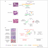
(a) Stromal area codes regulate leukocyte accumulation, differentiation and survival in the thymus, bone marrow and lymph node. Homeostatic chemokines (CXCL12, CXCL13, CCL19, CCL21), adhesion molecules [vascular cell adhesion molecule-1 (VCAM-1)] and cytokines and growth factors [interleukin-6 (IL-6), IL-7, fibroblast growth factor-7 (FGF-7), FGF-10] are components of the stromal code that help define stromal niches (bone marrow, thymus and lymph node) involved in leukocyte accumulation, differentiation and survival. Fibroblasts produce the appropriate cytokine or chemokine and express the appropriate adhesion receptor that is recognized by cognate receptors on the infiltrating leukocytes. In the case of the developing lymph node, ‘inducer’ lymphocytes produce lymphotoxin-α and IL-7, which induce the secretion of constitutive chemokines from the stromal ‘organizer’ cells, leading to the development of lymphoid aggregates with a lymph node structure. (b) Components of the stromal area code are aberrantly expressed in disease. During physiological inflammation, inflammatory chemokines (CCL2-CCL5, CX3CL1 and CXCL1-CXCL11) and inflammatory mediators, such as interferon-γ (IFN-γ), tumor necrosis factor-α (TNF-α) and IL-1, are produced by stromal cells and lead to the recruitment of inflammatory cells (lymphocytes, neutrophils and monocytes). During the resolution phase of inflammation, these proinflammatory stimuli are removed. However, in chronic persistent inflammation (pathological inflammation), stromal cells in tissues, such as the synovium, salivary gland, thyroid and liver, begin to aberrantly produce or express components of the physiological stromal code normally associated with primary and secondary lymphoid tissue, leading to the generation of pathogenic tertiary lymphoid tissue.

Similar articles

Cited by

References

    1. Campbell DJ, et al. Chemokines in the systemic organization of immunity. Immunol. Rev. 2003;195:58–71. - PubMed
    1. Warnock RA, et al. Molecular mechanisms of lymphocyte homing to peripheral lymph nodes. J. Exp. Med. 1998;187:205–216. - PMC - PubMed
    1. Nakano H, et al. A novel mutant gene involved in T-lymphocyte-specific homing into peripheral lymphoid organs on mouse chromosome 4. Blood. 1998;91:2886–2895. - PubMed
    1. Nakano H, et al. Genetic defect in T lymphocyte-specific homing into peripheral lymph nodes. Eur. J. Immunol. 1997;27:215–221. - PubMed
    1. Gunn MD, et al. Mice lacking expression of secondary lymphoid organ chemokine have defects in lymphocyte homing and dendritic cell localization. J. Exp. Med. 1999;189:451–460. - PMC - PubMed

MeSH terms

LinkOut - more resources