Comparative Study
. 2005 Mar 2;25(9):2245-54.
doi: 10.1523/JNEUROSCI.4956-04.2005.

Interactions between ephrin-B and metabotropic glutamate 1 receptors in brain tissue and cultured neurons

Affiliations
Comparative Study

Interactions between ephrin-B and metabotropic glutamate 1 receptors in brain tissue and cultured neurons

L Calò et al. J Neurosci. .

Abstract

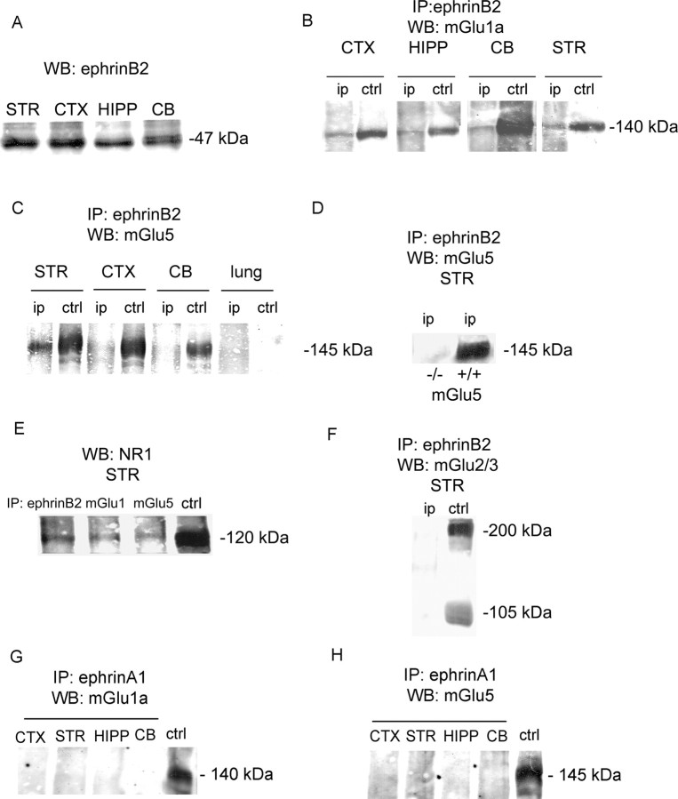
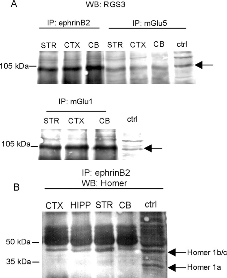
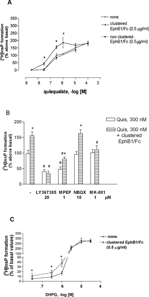
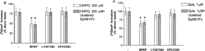
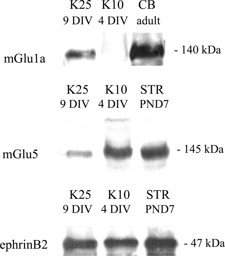
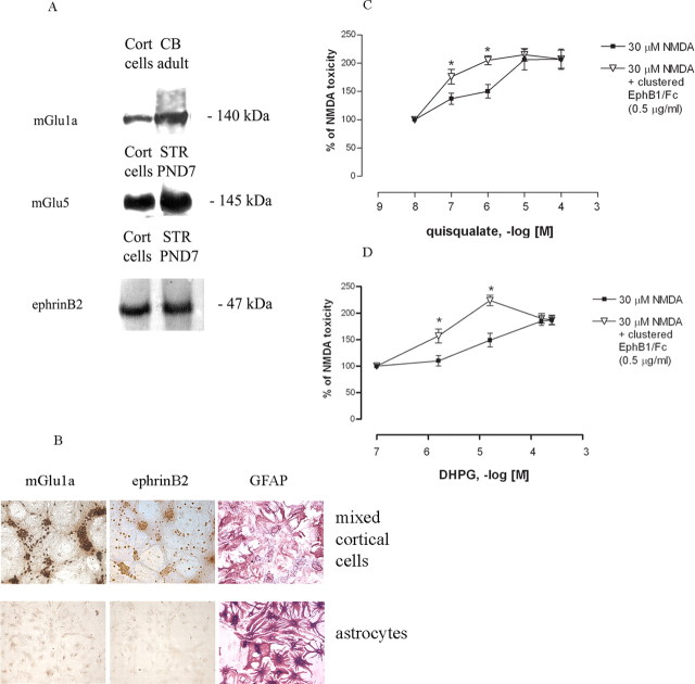
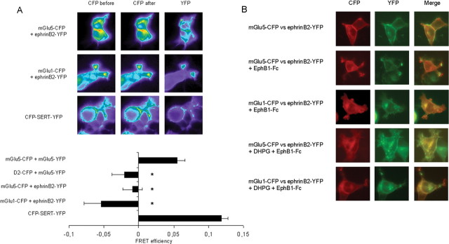
We examined the interaction between ephrins and metabotropic glutamate (mGlu) receptors in the developing brain and cultured neurons. EphrinB2 coimmunoprecipitated with mGlu1a receptors, in all of the brain regions examined, and with mGlu5 receptors in the corpus striatum. In striatal slices, activation of ephrinB2 by a clustered form of its target receptor, EphB1, amplified the mGlu receptor-mediated stimulation of polyphosphoinositide (PI) hydrolysis. This effect was abolished in slices treated with mGlu1 or NMDA receptor antagonists but was not affected by pharmacological blockade of mGlu5 receptors. An interaction among ephrinB2, mGlu1 receptor, and NMDA was supported by the following observations: (1) the NR1 subunit of NMDA receptors coimmunoprecipitated with mGlu1a receptors and ephrinB2 in striatal lysates; (2) clustered EphB1 amplified excitatory amino acid-stimulated PI hydrolysis in cultured granule cells grown under conditions that favored the expression of mGlu1a receptors; and (3) clustered EphB1 amplified the enhancing effect of mGlu receptor agonists on NMDA toxicity in cortical cultures, and its action was sensitive to mGlu1 receptor antagonists. Finally, fluorescence resonance energy transfer and coclustering analysis in human embryonic kidney 293 cells excluded a physical interaction between ephrinB2 and mGlu1a (or mGlu5 receptors). A functional interaction between ephrinB and mGlu1 receptors, which likely involves adaptor or scaffolding proteins, might have an important role in the regulation of developmental plasticity.


Figures

Figure 1.
Figure 1.
EphrinB2 expression and coimmunoprecipitation with group I mGlu receptors and NR1 subunit of NMDA receptors in the postnatal brain. Western blot analysis of ephrinB2 in the corpus striatum (STR), cerebral cortex (CTX), hippocampus (HIPP), and cerebellum (CB) is shown in A. B, C, Protein lysates of the CTX, HIPP, CB, and STR from P6-7 rats were immunoprecipitated (IP) with anti-ephrinB2 antibodies and then immunoblotted (WB) with anti-mGlu1a (B) or anti-mGlu5 (C) antibodies. Controls (ctrl) represent nonimmunoprecipitated samples. C, Extracts from the adult rat lung are shown as a negative control. Coimmunoprecipitation of ephrinB2 and mGlu5 receptors in the striatum of wild-type and mGlu5 knock-out mice at P6 is shown in D. E, Lysates from the striatum of P6-7 rats were immunoprecipitated with anti-ephrinB2, anti-mGlu1a, or anti-mGlu5 antibody and immunoblotted with anti-NR1 antibodies; the lack of coimmunoprecipitation between ephrinB2 and mGlu2/3 receptors in the striatum from P6-7 rats is shown in F. G, H, Neither mGlu1a nor mGlu5 receptors are detected in ephrinA1 immunoprecipitates of the CTX, STR, HIPP, and CB from rats at P6-7. Nonimmunoprecipitated cerebellum (G) and cerebral cortex (H) are used as a control (ctrl).
Figure 2.
Figure 2.
EphrinB2 coimmunoprecipitation with RGS3 and Homer in the postnatal brain. A, RGS3 is detected in ephrinB2, mGlu1a, and mGlu1a immunoprecipitates (IP) from P6-7 rats. The detection of Homer proteins in ephrinB2 immunoprecipitates is shown in B. All Homer proteins are simultaneously detected with a pan-Homer antibody. Note that the immunoreactive band corresponding to Homer 1b/c (but not that corresponding to Homer 1a) is detected in all ephrinB2 immunoprecipitates. CB, Cerebellum; ctrl, control; CTX, cerebral cortex; HIPP, hippocampus; STR, corpus striatum.
Figure 3.
Figure 3.
Activation of ephrinB by clustered EphB1/Fc amplifies the stimulation of PI hydrolysis by mGlu receptor agonists in striatal slices from P6-7 rats. A concentration-dependent stimulation of PI hydrolysis by quisqualate or DHPG in the absence or presence of clustered EphB1/Fc is shown in A and C, respectively. Figure 2 B shows that the enhancing effect of clustered EphB/Fc is abrogated by the mGlu1 receptor antagonist LY367385 or by the NMDA receptor antagonist MK-801 but not by the mGlu5 receptor antagonist MPEP or the AMPA receptor antagonist NBQX. Values are means ± SEM of 9-21 (A) or 6-12 (B, C) determinations; *p < 0.05 (Student's t test) compared with the respective values obtained in the absence of clustered EphB1/Fc; #p < 0.05 (one-way ANOVA plus Fisher's PLSD) compared with the respective values obtained with quisqualate or quisqualate plus clustered EphB2/Fc without receptor antagonists.
Figure 4.
Figure 4.
Clustered EphB1/Fc fails to amplify mGlu receptor-mediated PI hydrolysis in cortical slices from P6-7 rats. Note that the PI response to quisqualate (A) or DHPG (B) was attenuated by MPEP but not by the mGlu1 receptor antagonists CPCCOEt and LY367385. Values are means ± SEM of 12 determinations. #p < 0.05 (one-way ANOVA plus Fisher's PLSD) compared with the respective values obtained with quisqualate or quisqualate plus clustered EphB2/Fc without receptor antagonists.
Figure 5.
Figure 5.
Western blot analysis of mGlu1a receptor, mGlu5 receptors, and ephrinB2 in cultured cerebellar granule cells grown in medium containing 25 mm K+ (K25) at 9 DIV or in medium containing 10 mm K+ (K10) at 4 DIV is shown. Expression in the rat cerebellum (CB) or striatum (STR) is also shown.
Figure 6.
Figure 6.
Clustered EphB1/Fc amplifies the enhancing effect of group I mGlu receptor agonists on NMDA toxicity in cultured cortical cells. Western blot analysis of mGlu1a receptor, mGlu5 receptor, and ephrinB2 in mixed cultures of mouse cortical cells (Cort cells) is shown in A. Expression in the cerebellum (CB) and corpus striatum (STR) is also shown. Immunohistochemical analysis of mGlu1a receptors and ephrinB2 in mixed cultures of cortical cells (top) and in pure cultures of cortical astrocytes (bottom) are shown in B. Astrocytes in both cultures are stained with GFAP antibodies. Potentiation of NMDA toxicity by quisqualate or DHPG in cultured cortical cells incubated in the absence or presence of clustered EphB1/Fc is shown in B and C, respectively. Values are means ± 9 determinations from three independent experiments. *p < 0.05 (Student's t test) compared with the corresponding values obtained in the absence of clustered EphB1/Fc.
Figure 7.
Figure 7.
Lack of physical association between recombinantly expressed ephrinB2 and group I mGlu receptors. A, Donor photobleaching and DRAP FRET microscopy. The indicated pairs of CFP- or YFP-labeled constructs were coexpressed in HEK293 cells at 1:1 ratio of donor to acceptor. A CFP-labeled D2 dopamine receptor was used as a negative control. A double-labeled human serotonin transporter (SERT), which is known to form oligomers (Just et al., 2004), was used as a positive control. DRAP FRET microscopy experiments were performed 24 h after transfection. To measure DRAP, a donor (CFP) image was acquired before and after photobleaching using the YFP setting for 90 s (excitation, 500 nm; dichroic mirror, 525 nm; emission, 535 nm). FRET efficiencies (mean ± SE; calculated as described in Materials and Methods) were significantly different (p < 0.05, judged by one-way ANOVA followed by Tukey's multiple comparison test) from positive controls (mGlu5-CFP vs mGlu5-YFP; CFP-SERT-YFP), indicated by an asterisk. B, Clustering of YFP-tagged ephrinB2. Twenty-four hours after transfection, cells expressing both constructs were incubated at 37°C with EphB1-Fc preclustered with anti-human IgG according to the protocol described in Materials and Methods (in the presence or absence of DHPG; 50 μm). After 20 min, the coverslips were mounted for fluorescence microscopy, and images with CFP and YFP settings were acquired. The images are representative of three independent experiments.

Similar articles

Cited by

References

    1. Alagarsamy S, Marino MJ, Rouse ST, Gereau IV RW, Heinemann SF, Conn PJ (1999) Activation of NMDA receptors reverses desensitization of mGluR5 in native and recombinant systems. Nat Neurosci 2: 234-240. - PubMed
    1. Ango F, Pin JP, Tu JC, Xiao B, Worley PF, Bockaert J, Fagni L (2000) Dendritic and axonal targeting of type 5 metabotropic glutamate receptor is regulated by homer1 proteins and neuronal excitation. J Neurosci 20: 8710-8716. - PMC - PubMed
    1. Ango F, Robbe D, Tu JC, Xiao B, Worley PF, Pin JP, Bockaert J, Fagni L (2002) Homer-dependent cell surface expression of metabotropic glutamate receptor type 5 in neurons. Mol Cell Neurosci 20: 323-329. - PubMed
    1. Aniksztejn L, Sciancalepore M, Ben Ari Y, Cherubini E (1995) Persistent current oscillations produced by activation of metabotropic glutamate receptors in immature rat CA3 hippocampal neurons. J Neurophysiol 73: 1422-1429. - PubMed
    1. Awad H, Hubert GW, Smith Y, Levey AI, Conn PJ (2000) Activation of metabotropic glutamate receptor 5 has direct excitatory effects and potentiates NMDA receptor currents in neurons of the subthalamic nucleus. J Neurosci 20: 7871-7879. - PMC - PubMed

Publication types

MeSH terms