Comparative Study
. 2005 Mar 2;25(9):2285-94.
doi: 10.1523/JNEUROSCI.3877-04.2005.

Neurosteroid-induced plasticity of immature synapses via retrograde modulation of presynaptic NMDA receptors

Affiliations
Comparative Study

Neurosteroid-induced plasticity of immature synapses via retrograde modulation of presynaptic NMDA receptors

Manuel Mameli et al. J Neurosci. .

Abstract

Neurosteroids are produced de novo in neuronal and glial cells, which begin to express steroidogenic enzymes early in development. Studies suggest that neurosteroids may play important roles in neuronal circuit maturation via autocrine and/or paracrine actions. However, the mechanism of action of these agents is not fully understood. We report here that the excitatory neurosteroid pregnenolone sulfate induces a long-lasting strengthening of AMPA receptor-mediated synaptic transmission in rat hippocampal neurons during a restricted developmental period. Using the acute hippocampal slice preparation and patch-clamp electrophysiological techniques, we found that pregnenolone sulfate increases the frequency of AMPA-mediated miniature excitatory postsynaptic currents in CA1 pyramidal neurons. This effect could not be observed in slices from rats older than postnatal day 5. The mechanism of action of pregnenolone sulfate involved a short-term increase in the probability of glutamate release, and this effect is likely mediated by presynaptic NMDA receptors containing the NR2D subunit, which is transiently expressed in the hippocampus. The increase in glutamate release triggered a long-term enhancement of AMPA receptor function that requires activation of postsynaptic NMDA receptors containing NR2B subunits. Importantly, synaptic strengthening could also be triggered by postsynaptic neuron depolarization, and an anti-pregnenolone sulfate antibody scavenger blocked this effect. This finding indicates that a pregnenolone sulfate-like neurosteroid is a previously unrecognized retrograde messenger that is released in an activity-dependent manner during development.


Figures

Figure 4.
Figure 4.
The PREGS-induced long-term enhancement of postsynaptic AMPA responses in P3-P4 slices is NMDA receptor and Ca2+ dependent. a1, Traces illustrating that the late phase of the 25 μm PREGS-induced increase of AMPA mEPSC frequency cannot be observed when the Ca2+ chelator BAPTA (10 mm) is internally dialyzed via the patch electrode. Calibration: 15 pA, 100 ms. a2, time course of the effect of 25 μm PREGS on a neuron internally dialyzed with BAPTA and a control neuron from the same batch of slices. a3, Summary of the effect of 25 μm PREGS on neurons internally dialyzed with 10 mm BAPTA (**p < 0.01; ***p < 0.001; n = 6). b1, NMDA receptor-mediated EPSCs (Vhold = +40 mm; calibration: 20 pA, 200 ms) are significantly inhibited by intracellular dialysis of MK-801 (5 mm; see Results for more details; ***p < 0.001; n = 11 by t test). To promote entrance of MK-801 to the pore, neurons were depolarized from -70 to -15 mV (5 times for 10 s each). b2, In six of these 11 neurons, after NMDA EPSC inhibition was confirmed, 500 nm tetrodotoxin was applied, and the effect of 25 μm PREGS on mEPSC frequency was assessed [experiment (Exp.) 1]. Under these conditions, application of PREGS did not induce a long-lasting increase in mEPSC frequency. In a separate batch of neurons, tetrodotoxin was present from the start of the experiment, and basal mEPSC frequency was recorded (experiment 2; see Results for more details). Neurons were then depolarized as described above to promote entrance of MK-801 to the pore. mEPSC frequency was recorded again and found to be unchanged by the depolarization procedure (see Results). As shown in the summary graph, 25 μm PREGS did not induce a long-lasting increase in mEPSC frequency under these conditions (n = 4). *p < 0.05; **p < 0.01; ***p < 0.001. c1, Extracellular application of 10 μm ifenprodil (ifen) induces a long-lasting decrease in the amplitude of NMDA EPSCs (recorded at Vhold = -10 mV). Note that these events are blocked by 100 μm dl-AP-5. Calibration: 20 pA, 200 ms. c2, In a separate batch of neurons, mEPSC frequency was recorded in the presence of tetrodotoxin (500 nm). Under these conditions, ifenprodil blocked the late phase of the 25 μm PREGS effect on mEPSC frequency. Ifenprodil alone did not have an effect on mEPSC frequency (see Results). *p < 0.05; **p < 0.01; n = 6. Ctrl, Control. Error bars represent SEM.
Figure 5.
Figure 5.
The PREGS effect in P3-P4 slices requires influx of Ca2+ through presynaptic NMDA receptors likely containing NR2D subunits. a, Preincubation with the membrane-permeable Ca2+ chelator BAPTA-AM (50 μm) blocks the early and late phases of the 25 μm PREGS-induced increase of mEPSC frequency (n = 3 for control and n = 4 for BAPTA-AM). b, A similar effect was observed in slices incubated with the nonselective NMDA receptor antagonists dl-APV (100 μm; n = 3 for control and n = 5 for dl-APV) and 7-chlorokynurenate (7-CLKA; 30 μm; n = 5) and the antagonist of NMDA receptors containing NR2D subunits, PPDA (0.1 μm; n = 5). c1, Bath application of the NMDA receptor agonist homoquinolinic acid (HQA; 10 μm) induces an inward current in a P4 neuron that is blocked by intracellular dialysis of MK-801 (5 mm). Calibration: 100 pA, 300 s. c2, Sample traces illustrating that, in the presence of internal MK-801, homoquinolinic acid mimics the PREGS-induced increase of mEPSC frequency in slices from P3 but not P8 rats. Calibration: 20 pA, 300 ms. c3, Summary graph illustrating the effect of homoquinolinic acid, in the presence of internal MK-801, on P3-P4 (n = 6). Lack of an effect of homoquinolinic acid on P6-P10 slices (n = 5) and blockade of its effect by PPDA (0.1 μm) in P3-P4 slices (n = 5) are also shown. *p < 0.05; **p < 0.01; ***p < 0.001. Ctrl, Control. Error bars represent SEM.
Figure 1.
Figure 1.
PREGS persistently increases mEPSC frequency in immature synapses in an age-dependent and concentration-dependent manner. a1, Brief (5 min) exposure to 25 μm PREGS persistently increases mEPSC frequency in a P3 CA1 pyramidal neuron. Calibration: 10 pA, 100 ms. a2, Cumulative probability plots for interevent interval (IEI) and amplitude corresponding to the recording shown above. Note the shift to the left in the IEI plot observed after a 20 min washout period after brief exposure to 25 μm PREGS. a3, Time course of the effect of brief (5 min) exposure to 17 μm (n = 4) and 25 μm (n = 8) PREGS on mEPSC frequency (*p < 0.05; **p < 0.01) and amplitude (error bars are smaller than the symbols) in P3-P4 neurons. b1, PREGS does not affect mEPSC frequency in a P7 neuron. Calibration: 10 pA, 100 ms. b2, PREGS increases mEPSC frequency only in P3-P5 neurons (**p < 0.01 and ***p < 0.001 vs P20 neurons; n = 4-12). KCl (10 mm in ACSF) increases mEPSC frequency to a similar extent in P3-P4 and P7 neurons (n = 4). c, The effect of 25 μm PREGS is independent of basal mEPSC frequency. Note that the range of basal mEPSC frequency is similar in slices from P3-P4 and P6-P10 rats. d1, Traces illustrating the effect of increasing PREGS concentrations on mEPSC frequency in P3-P4 neurons. Calibration: 10 pA, 500 ms. d2, PREGS increases mEPSC frequency at concentrations between 17 and 50 μm (*p < 0.05 and ***p < 0.001; n = 4-9). Note the lack of an effect of vehicle (0.025% Me2SO, which was the concentration delivered to the cells in the experiments with 50 μm PREGS). Ctrl, Control. Error bars represent SEM.
Figure 2.
Figure 2.
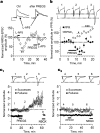
PREGS transiently increases glutamate release and triggers a long-term enhancement of AMPA receptor-mediated synaptic responses in P3-P4 slices. a, Evoked NMDA EPSCs are not inhibited by the weak antagonist l-AP-5 (250 μm) after exposure to 25 μm PREGS because of increased glutamate release probability. Calibration: 20 pA, 200 ms (**p < 0.01; n = 4). b, Time course of the effect of PREGS on the amplitude of the first EPSC (EPSC1) and paired-pulse ratio (PPR; 50 ms interpulse interval; stimulation delivered every 20 s). Calibration: 20 pA, 25 ms (*p < 0.05, **p < 0.01, and ***p < 0.001; n = 8). c1, Time course of the effect of 25 μm PREGS on AMPA EPSC failures and successes (n = 9). c2, Time course illustrating the lack of an effect of vehicle (0.012% Me2SO) on AMPA EPSC failures and successes (n = 9). NBQX (10 μm) was applied at the end of the recording to confirm that responses were mediated by AMPA receptors. Calibration: 27 pA, 15 ms. The bin size for the data shown in c1 and c2 is 1 min (stimulation was delivered every 20 s), and successes were defined as events with amplitude ≥4 pA. Amplitude of both successes and failures was normalized with respect to the average amplitude of successes obtained during the first 3 min of recording. Ctrl, Control. Error bars represent SEM.
Figure 3.
Figure 3.
PREGS induces a delayed potentiation of postsynaptic AMPA receptors. a, Effect of vehicle (0.012% Me2SO) and 25 μm PREGS on currents evoked by bath application of AMPA for 30 s (represented by the dots) in the presence of cyclothiazide (30 μm) and tetrodotoxin (500 nm). Currents were blocked by the AMPA receptor antagonist GYKI-53655 (30 μm). Calibration: 200 pA, 800 s (**p < 0.01; n = 4). b, Effect of 25 μm PREGS on currents evoked by pressure application of AMPA (5 μm in a micropipette located ∼200 μm from the soma) in the presence of both tetrodotoxin (500 nm) and bicuculline (20 μm) and in the absence of cyclothiazide. Currents were blocked by the AMPA receptor antagonist NBQX (10 μm; data not shown). AMPA puffs of 500 ms duration were delivered every 40 s, and their onset is represented by the dots above the traces. The summary graph shows average amplitudes in bins of 5 min. Amplitude was normalized with respect to the average amplitude of the responses obtained during the first 3 min of recording. Calibration: 40 pA, 500 ms (**p < 0.01, ***p < 0.001; n = 4). Error bars represent SEM.
Figure 6.
Figure 6.
A PREGS-like neurosteroid, which retrogradely modulates NMDA receptors, mediates the depolarization-induced increase of mEPSC frequency. a, Depolarization (depol.)-induced increase of mEPSC frequency can be observed in CA1 pyramidal neurons from P3-P4 (n = 19) but not P6-P10 (n = 7; traces not shown) rats. The cumulative probability plots and average traces (inset) illustrate that mEPSC amplitude also increased 20 min after depolarization in P3-P4 neurons (compare solid vs dashed traces). A similar result was obtained with P6-P10 neurons (n = 7; data not shown). b, Preincubation (14 min) with rabbit anti-PREGS IgG blocks the depolarization-induced increase of mEPSC frequency but not the increase in amplitude in P3-P4 neurons (n = 14). c, Incubation with rabbit IgG neither affects the depolarization-induced increase in frequency nor the increase in amplitude in P3-P4 neurons (n = 6). d, Incubation with PPDA blocks the depolarization-induced increase of mEPSC frequency but not the increase in amplitude in P3-P4 neurons (n = 5). Calibration: a-d, 20 pA, 5 s (left); 8 pA, 12 ms (right). *p < 0.05; **p < 0.01; ***p < 0.001. Ctrl, Control. Error bars represent SEM.

Similar articles

Cited by

References

    1. Banke TG, Bowie D, Lee H, Huganir RL, Schousboe A, Traynelis SF (2000) Control of GluR1 AMPA receptor function by cAMP-dependent protein kinase. J Neurosci 20: 89-102. - PMC - PubMed
    1. Baulieu EE, Robel P, Schumacher M (2001) Neurosteroids: beginning of the story. Int Rev Neurobiol 46: 1-32. - PubMed
    1. Ben-Ari Y (2002) Excitatory actions of GABA during development: the nature of the nurture. Nat Rev Neurosci 3: 728-739. - PubMed
    1. Berretta N, Jones RS (1996) Tonic facilitation of glutamate release by presynaptic N-methyl-d-aspartate autoreceptors in the entorhinal cortex. Neuroscience 75: 339-344. - PubMed
    1. Blake JF, Brown MW, Collingridge GL (1988) A quantitative study of the actions of excitatory amino acids and antagonists in rat hippocampal slices. Br J Pharmacol 95: 291-299. - PMC - PubMed

Publication types

MeSH terms

LinkOut - more resources