Comparative Study
. 2005 Mar 2;25(9):2434-44.
doi: 10.1523/JNEUROSCI.4517-04.2005.

Coxsackievirus targets proliferating neuronal progenitor cells in the neonatal CNS

Affiliations
Comparative Study

Coxsackievirus targets proliferating neuronal progenitor cells in the neonatal CNS

Ralph Feuer et al. J Neurosci. .

Abstract

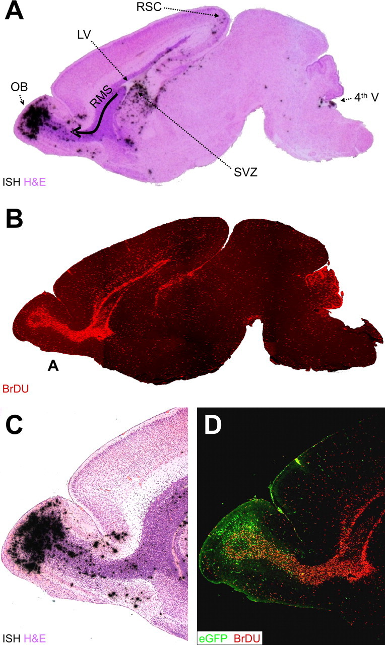
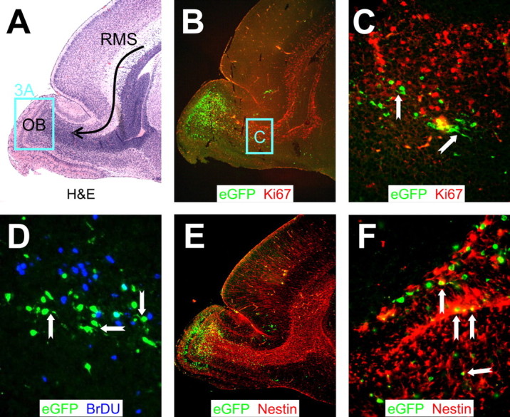
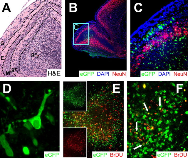
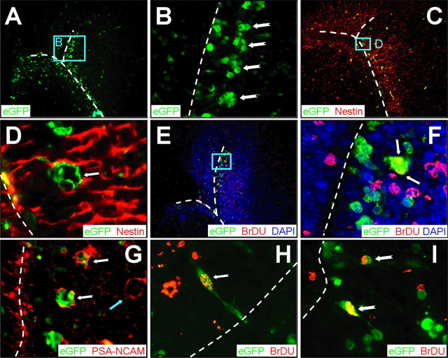
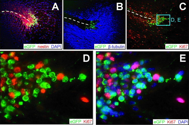
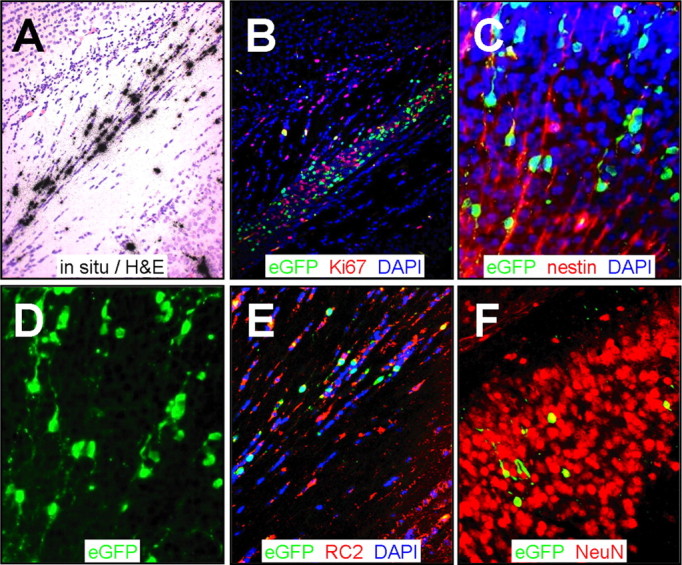
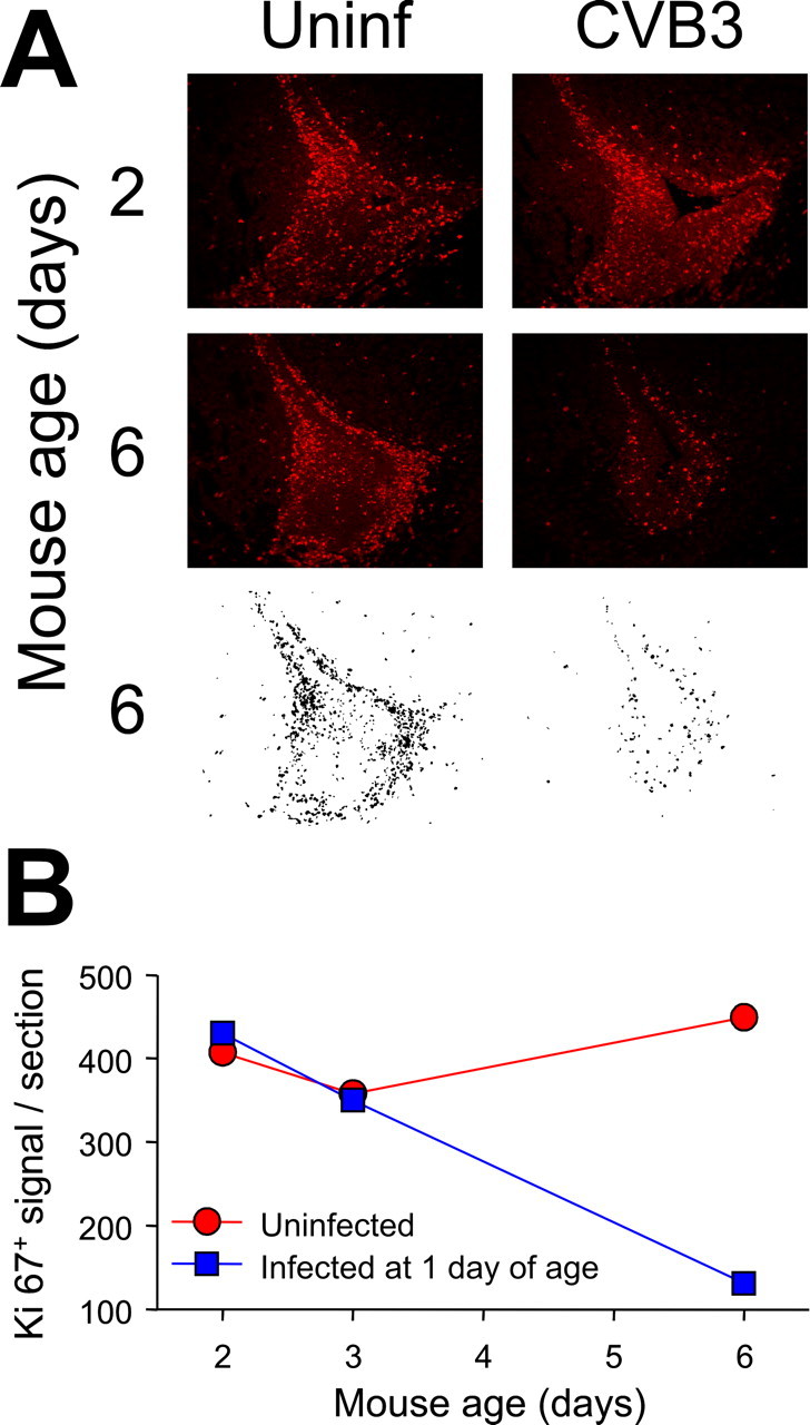
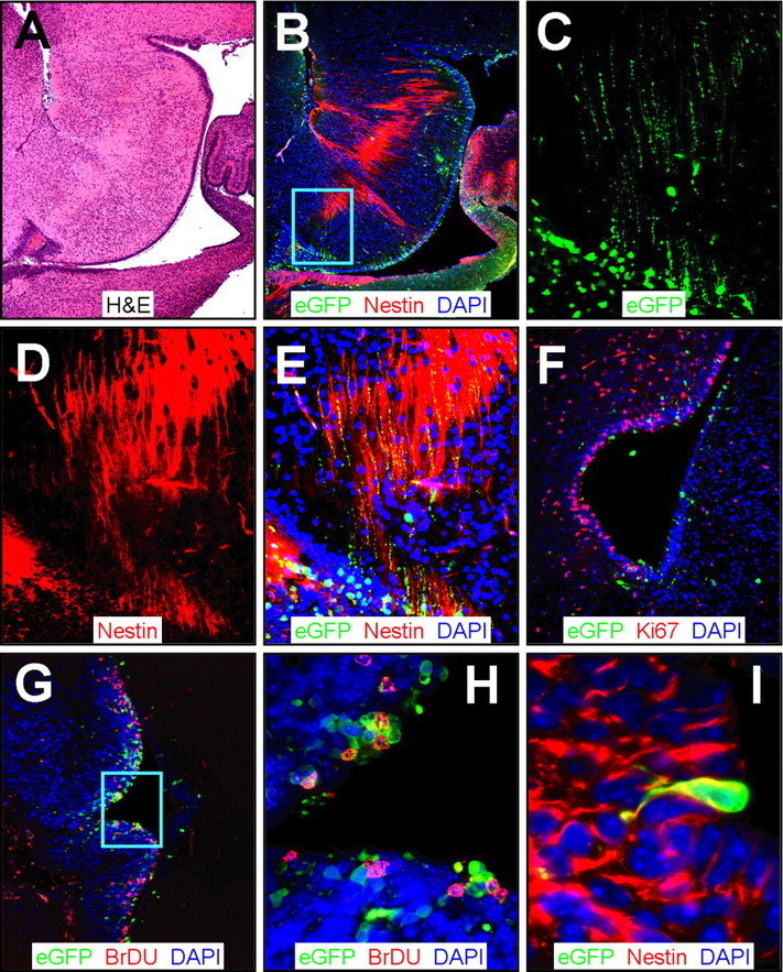
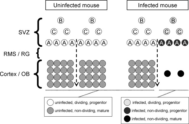
Type B coxsackieviruses (CVB) frequently infect the CNS and, together with other enteroviruses, are the most common cause of viral meningitis in humans. Newborn infants are particularly vulnerable, and CVB also can infect the fetus, leading to mortality, or to neurodevelopmental defects in surviving infants. Using a mouse model of neonatal CVB infection, we previously demonstrated that coxsackievirus B3 (CVB3) could infect neuronal progenitor cells in the subventricular zone (SVZ). Here we extend these findings, and we show that CVB3 targets actively proliferating (bromodeoxyuridine+, Ki67+) cells in the SVZ, including type B and type A stem cells. However, infected cells exiting the SVZ have lost their proliferative capacity, in contrast to their uninfected companions. Despite being proliferation deficient, the infected neuronal precursors could migrate along the rostral migratory stream and radial glia, to reach their final destinations in the olfactory bulb or cerebral cortex. Furthermore, infection did not prevent cell differentiation, as determined by cellular morphology and the expression of maturation markers. These data lead us to propose a model of CVB infection of the developing CNS, which may explain the neurodevelopmental defects that result from fetal infection.


Figures

Figure 1.
Figure 1.
Distribution of CVB and proliferating cells in the neonatal CNS. We treated 1-d-old pups with BrDU (intraperitoneally), and we infected them with eGFP-CVB3 (2 × 10⁶ pfu, i.c.). After 2 d, the brain was harvested and fixed in 10% neutral-buffered Formalin. Sagittal sections were deparaffinized (see Materials and Methods) and probed for viral RNA by in situ hybridization or immunostained using antibody against BrDU. A, H&E-stained sagittal section, close to the midline, with viral RNA revealed by in situ hybridization (black dots). Viral RNA is present in the OB, the RMS (curved arrow), the ventral SVZ below the lateral ventricle (LV), the RSC, and in the region of the fourth ventricle (4th V). B, A similar sagittal section (composite of 5× images) showing BrDU staining (red) that highlights the RMS, as well as the cerebellar cortex. C, Higher-power image (5×) showing the layered distribution of viral RNA in the OB and scattered throughout the RMS. D, Viral protein expression is shown (5×) by eGFP (green), and cells that have divided are indicated by BrDU staining (red).
Figure 2.
Figure 2.
CVB3 protein expression in migratory neuroblasts within proliferating regions of the RMS. Sagittal sections of the same infected brain presented in Figure 1 were immunostained with antibodies against Ki67, BrDU, and nestin. A, H&E staining. The curved black arrow represents the RMS, which contains neuroblasts migrating from the SVZ to the OB. The distinct layers of the olfactory bulb were apparent, and a higher magnification of this area (represented by the cyan box) is evaluated in Figure 3A. B, Proliferating cells in the RMS were detected using an antibody against Ki67. Infected cells (eGFP+) were observed within proliferating (Ki67+; red) regions extending from the anterior SVZ into the olfactory bulb. C, Higher magnification (B, cyan box) revealed infected cells in close proximity to, yet distinct from, proliferating cells. Many infected cells in the RMS exhibited morphological similarities to migratory neuroblasts, with characteristic leading or lagging axonal progressions (notched arrows). D, Similarly, infected cells in the RMS were observed in close proximity to, yet distinct from, BrDU+ (blue) cells. Notched arrows indicate infected migratory neuroblasts. E, Nestin+ (red) staining was seen throughout the RMS. F, Higher magnification of E demonstrated direct colocalization of nestin with infected migratory neuroblasts. A, B, E, 5× objective; C, 20× objective with an additional twofold computer-generated magnification; D, 20× objective with an additional threefold computer-generated magnification; F, 40× objective.
Figure 3.
Figure 3.
CVB3 protein expression and markers of cellular proliferation and maturation in the OB. All samples represent brains from mice that were infected at 1 d after birth and killed 2 d after infection. A, High magnification of the OB region shown in Figure 2A. The various layers of the OB, described in detail in Results, are separated by dashed black lines. B, Infected cells positive for NeuN were identified in the grs cell layer of the OB (5× magnification). The majority of NeuN staining (red) included a distinct halo of cells extending within the grs layer of the olfactory bulb and continuing throughout the cortex. Very little NeuN staining was identified within the RMS. DAPI stain (blue), which identified the nucleus of all cells, helped to define tissue section morphology. DAPI signal merged with NeuN generated a magenta color illustrating the nuclear localization of NeuN antigen. C, Higher magnification (B, cyan box) of the olfactory bulb demonstrated infection in the mitral cell layer and in granule cells in both the grs and grd layers. Infected cells in the mitral cell layer were NeuN-. However, infected granule cells were NeuN+ in the grs cell layer and NeuN- in the grd cell layer of the OB. The periglomerular cell layer was spared from infection. D, Many infected cells in the mitral cell layer had characteristic mitral cell morphology with a single projection positioned toward the periglomerular cell layer. E, Regional colocalization of virus protein expression (eGFP+) and cellular proliferation (BrDU+; red) was observed in the granule cell layer of the olfactory bulb. Individual channels for eGFP or BrDU (insets) demonstrated a close association of infected and dividing cells. F, Higher magnification of the olfactory bulb revealed some BrDU+ infected cells (yellow; notched arrows). A, C, E, 20× objective; B, 5× objective; D, 100.8× objective; F, 63× objective.
Figure 4.
Figure 4.
CVB3 infects type A, as well as type B, stem cells. Transverse paraffin-embedded sections were deparaffinized and immunostained using antibodies against nestin, BrDU, or PSA-NCAM. The ependymal cell layer is represented by the dashed white line. A, Many clusters of infected cells (eGFP+) were observed within the subventricular zone. B, Higher magnification (A, cyan box) revealed the dense clusters of infected cells (notched arrows) morphologically similar to type A neuronal progenitor cells within the SVZ. C, As expected, nestin staining (red) was observed near the lateral ventricle and within the SVZ. D, Higher magnification (C, cyan box) demonstrated that these clusters of infected cells were nestin+, suggesting that these cells share immunological similarities with type A or type C neuronal progenitor cells. E, BrDU staining (red) indicated that many cells near the SVZ had recently divided. F, Higher magnification (E, cyan box) showed direct colocalization between infected and BrDU+ cells (arrows), indicating that these clusters of cells had recently divided. G, PSA-NCAM staining (red) allowed us to distinguish type A and type C neuronal progenitor cells in the SVZ. Clusters of infected cells (notched white arrows) near the SVZ were found to be PSA-NCAM+, which suggests that type A neuronal progenitor cells are susceptible to infection; uninfected clusters also were present (notched cyan arrow). H, Merged images revealed colocalization of nuclear BrDU+ staining (red) and infected cells (cytoplasmic eGFP) near the lateral ventricle. An infected cell (100× with an additional twofold computer-generated magnification) labeled with BrDU (notched arrow) is visible protruding through the ependymal cell layer into the ventricle, having morphological similarities to a type B cell. I, Two infected cells labeled with BrDU are shown deeper within the subventricular zone. A, C, E, 20× objective; B, 100× objective; D, F, H, I, 100× objective with an additional twofold computer-generated magnification; G, 20× objective with an additional threefold computer-generated magnification.
Figure 5.
Figure 5.
Proliferation and infection are mutually exclusive in cells exiting the SVZ. Transverse paraffin-embedded sections from an infected brain 1 d after infection were immunostained using antibodies against nestin, β-tubulin, and Ki67. Dashed lines represent the ependymal cell layer of the lateral ventricle. A, Nestin staining (red) colocalized with the majority of infected (eGFP+) cells in the SVZ. B, The majority of β-tubulin expression (blue) was detected farther away from the SVZ, although a few infected cells were observed within the β-tubulin staining region. C, Infected cells regionally colocalized with proliferating cells expressing Ki67 (red) in the subventricular zone near the lateral ventricle. D, Higher magnification (C, cyan box) revealed infected cells adjacent to, yet distinct from, Ki67+ cells. Almost all Ki67+ cells were eGFP-, and nearly all eGFP+ cells were Ki67-. E, DAPI (blue) nuclear counterstain allowed visualization of all cells within the section and illustrated the lack of Ki67/eGFP colocalization. The nuclear localization of Ki67 antigen and cytosolic distribution of eGFP were evident after merging the red, blue, and green channels. A-C, 20× objective; D, E, 100.8× objective.
Figure 7.
Figure 7.
Virus-infected cells are arrayed as chains, contiguous with radial glia. Paraffin-embedded sections were obtained from the brain 2 d after infection and stained using antibodies against Ki67, nestin, and RC2. Viral infection was evaluated by in situ hybridization (CVB3 5′ untranslated region probe) or by viral protein expression (eGFP). A, In situ hybridization identified infected cells near the retrosplenial cortex in longitudinal arrays. B, Proliferating cells were identified in close proximity to infected cells (eGFP+) in this region. C, Sections stained for nestin and counterstained with DAPI (blue) revealed infected nestin+ cells that appeared to be migratory neuroblasts associated with radial glia. D, Single-channel (eGFP) image of C highlighted the chain-like distribution of infected cells with long extensions entering into the cortex, reminiscent of radial glial cells. E, RC2 staining (red) identified many infected radial glial cells contiguous with the retrosplenial cortex. F, Many infected NeuN+ cells were observed deeper within the cortex. A, B, 20× objective; C, D, 20× objective with an additional approximately threefold computer-generated magnification; E, F, 20× objective with an additional approximately two-fold computer-generated magnification.
Figure 6.
Figure 6.
Quantitative analysis of Ki67+ cells in the SVZ. A, We infected 1-d-old pups with eGFP-CVB3 (2 × 10⁶ pfu, i.c.), and they were killed 1, 2, or 5 d later. Uninfected pups of the same ages were evaluated for comparative purposes. Sections of the SVZ were stained with an antibody against Ki67 (red) and observed by fluorescence microscopy. To enumerate Ki67+ cells, black and white images were generated using ImageJ, as described in Materials and Methods; examples are shown for the 6-d-old mice. B, The number of Ki67+ cells per section are shown at each time point for uninfected mice (red) and infected mice (blue).
Figure 8.
Figure 8.
CVB3 infection adjacent to the fourth ventricle. Sagittal (A-E) and transverse (F-I) paraffin sections of the posterior brain of a newborn mouse at 1 d after infection were immunostained using antibodies against nestin, Ki67, and BrDU. A, Low-magnification H&E-stained sagittal section identified features of the cerebellum, the fourth ventricle, and the ependymal cell layer. B, Infected cells (eGFP+) were evident near the ependymal cell layer in the area below the fourth ventricle and in the region of the dorsal tegmental nucleus. Nestin+ staining (red) was observed stretching across the ependymal cell layer into the surrounding parenchyma. The cyan box represents the area expanded in C-E. C, Higher magnification revealed infected (eGFP+) cells with long cellular processes. D, Similarly, nestin staining demonstrated long extensions of neuronal progenitor cells found near the fourth ventricle. E, Direct colocalization of nestin and infected axonal processes was evident (yellow), indicating that neuronal progenitor cells in the fourth ventricle were susceptible to infection. F, Ki67 staining (red) showed many proliferating cells near the ependymal cell layer and infected regions of the fourth ventricle. G, Similarly, many BrDU cells (red) were found closely associated with infected cells near the ependymal cell layer. H, Higher magnification (G, cyan box) demonstrated direct colocalization of many infected cells with BrDU staining. I, Many nestin+ (red) infected cells protruded through the ependymal cell layer in the fourth ventricle, consistent with infected type B stem cells, as seen near the lateral ventricle (Fig. 4).
Figure 9.
Figure 9.
Model for CVB3 dissemination in the neonatal CNS. A graphical representation of CVB3 infection of the neonatal CNS. The SVZ of uninfected mice (left column) contains three populations of neuronal progenitor cells (white circles). Type B cells give rise to type C cells, which eventually produce migratory neuroblasts (type A cells). All progenitor cells are known to proliferate extensively. Neuronal progenitor cells migrate to the OB or cortex through the RMS or radialglia (RG) and differentiate into mature neurons (hatched circles). In an infected mouse (right column), CVB3 infects some of the type B and A progenitor cells, and type C cells also may be susceptible to infection. CVB3 inhibits the proliferation of neuronal progenitor cells exiting the SVZ (dark gray circles) as judged by Ki67 expression (quantitated in Fig. 6); these nondividing progenitor cells nevertheless retained their ability to migrate and to differentiate into infected mature neurons (black circles). However, many of these infected neurons may eventually die as a result of caspase-3-mediated apoptosis, and this, together with the reduction in Ki67+ cells exiting the SVZ, may lead to a marked reduction in mature neurons (right side of infected mouse column) and thereby to the neurodevelopmental defects associated with CVB infections.

Similar articles

Cited by

References

    1. Alvarez-Buylla A, Garcia-Verdugo JM, Tramontin AD (2001) A unified hypothesis on the lineage of neural stem cells. Nat Rev Neurosci 2: 287-293. - PubMed
    1. Baker H, Liu N, Chun HS, Saino S, Berlin R, Volpe B, Son JH (2001) Phenotypic differentiation during migration of dopaminergic progenitor cells to the olfactory bulb. J Neurosci 21: 8505-8513. - PMC - PubMed
    1. Daley AJ, Isaacs D, Dwyer DE, Gilbert GL (1998) A cluster of cases of neonatal coxsackievirus B meningitis and myocarditis. J Paediatr Child Health 34: 196-198. - PubMed
    1. Doetsch F (2003) A niche for adult neural stem cells. Curr Opin Genet Dev 13: 543-550. - PubMed
    1. Doetsch F, Garcia-Verdugo JM, Alvarez-Buylla A (1997) Cellular composition and three-dimensional organization of the subventricular germinal zone in the adult mammalian brain. J Neurosci 17: 5046-5061. - PMC - PubMed

Publication types

MeSH terms