

Wound healing in rat cornea: the role of electric currents
- PMID: 15746181
- PMCID: PMC1459277
- DOI: 10.1096/fj.04-2325com
Wound healing in rat cornea: the role of electric currents
Abstract
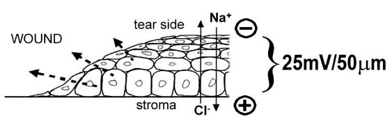
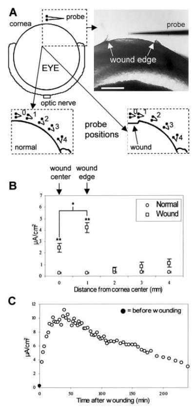
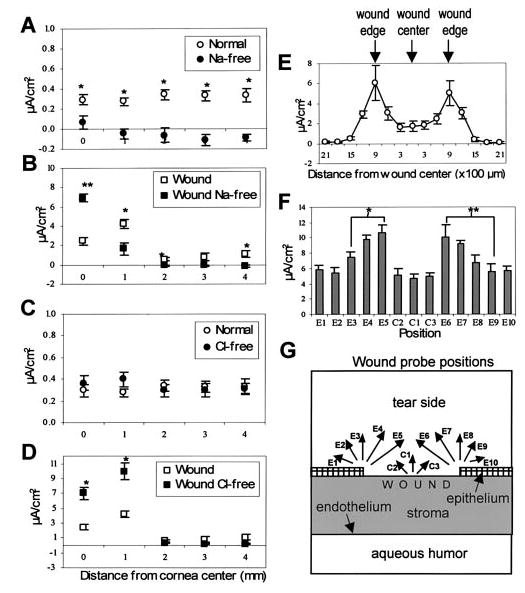
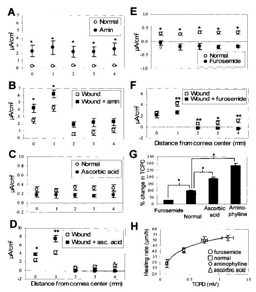
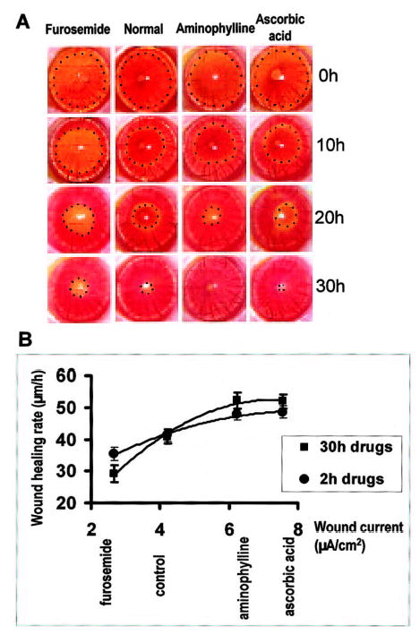
Human corneal epithelial cells respond rapidly following injury to restore the integrity of the ocular surface. What stimulates and guides cells to move into the wound to heal? One candidate is the wound-induced electric field. Using vibrating probe techniques, we provide detailed temporal and spatial mapping of endogenous electric currents at rat corneal wounds. We find Cl- and Na+ are the major components of electric currents in rat corneal wounds. Na+ is the major component of ionic transport in the resting (nonwounded) rat cornea and of the wound center leakage current, whereas Cl- is a more important component of the endogenous electrical current at the wound edges. Enhancing or decreasing Cl- flow with clinically approved pharmacological agents such as aminophylline, ascorbic acid, or furosemide increased or decreased endogenous wound electric currents, respectively. These changes in wound currents correlated directly with the rate of wound healing in vivo. Thus, pharmacologically enhancing or decreasing wound-induced electric currents increased and decreased wound healing rate, respectively. This may have wide-reaching and novel therapeutic potential in the management of wound healing and may help explain some mechanistic aspects of the effects of some clinically used agents.
Figures
Similar articles
-
Modulating endogenous electric currents in human corneal wounds--a novel approach of bioelectric stimulation without electrodes.Cornea. 2011 Mar;30(3):338-43. doi: 10.1097/ICO.0b013e3181f7f2de. Cornea. 2011. PMID: 21099404 Free PMC article.
-
Ionic components of electric current at rat corneal wounds.PLoS One. 2011 Feb 25;6(2):e17411. doi: 10.1371/journal.pone.0017411. PLoS One. 2011. PMID: 21364900 Free PMC article.
-
Electrical cues regulate the orientation and frequency of cell division and the rate of wound healing in vivo.Proc Natl Acad Sci U S A. 2002 Oct 15;99(21):13577-82. doi: 10.1073/pnas.202235299. Epub 2002 Oct 4. Proc Natl Acad Sci U S A. 2002. PMID: 12368473 Free PMC article.
-
Progress in corneal wound healing.Prog Retin Eye Res. 2015 Nov;49:17-45. doi: 10.1016/j.preteyeres.2015.07.002. Epub 2015 Jul 18. Prog Retin Eye Res. 2015. PMID: 26197361 Free PMC article. Review.
-
Thymosin beta4 and corneal wound healing: visions of the future.Ann N Y Acad Sci. 2010 Apr;1194:190-8. doi: 10.1111/j.1749-6632.2010.05472.x. Ann N Y Acad Sci. 2010. PMID: 20536468 Review.
Cited by
-
The role of transcription-independent damage signals in the initiation of epithelial wound healing.Nat Rev Mol Cell Biol. 2013 Apr;14(4):249-62. doi: 10.1038/nrm3541. Epub 2013 Feb 27. Nat Rev Mol Cell Biol. 2013. PMID: 23443750 Review.
-
A current affair: electrotherapy in wound healing.J Multidiscip Healthc. 2017 Apr 20;10:179-194. doi: 10.2147/JMDH.S127207. eCollection 2017. J Multidiscip Healthc. 2017. PMID: 28461755 Free PMC article. Review.
-
PI3K mediated electrotaxis of embryonic and adult neural progenitor cells in the presence of growth factors.Exp Neurol. 2011 Jan;227(1):210-7. doi: 10.1016/j.expneurol.2010.11.002. Epub 2010 Nov 16. Exp Neurol. 2011. PMID: 21092738 Free PMC article.
-
Transcutaneous Electrical Stimulation for the Prevention of Dry Eye Disease after Photorefractive Keratectomy: Randomized Controlled Trial.Ophthalmol Sci. 2022 Nov 2;3(2):100242. doi: 10.1016/j.xops.2022.100242. eCollection 2023 Jun. Ophthalmol Sci. 2022. PMID: 36685712 Free PMC article.
-
Infection-generated electric field in gut epithelium drives bidirectional migration of macrophages.PLoS Biol. 2019 Apr 9;17(4):e3000044. doi: 10.1371/journal.pbio.3000044. eCollection 2019 Apr. PLoS Biol. 2019. PMID: 30964858 Free PMC article.
References
-
- Clark, R. A. F., Ed (1996) The Molecular and Cellular Biology of Wound Repair, Plenum, New York
-
- Chiang M, Cragoe EJ, Vanable JW. Intrinsic electric fields promote epithelisation of wounds in the newt, Notophthalmus viridescens. Dev Biol. 1991;146:377–385. - PubMed
-
- Ojingwa JC, Isseroff RR. Electrical stimulation of wound healing. J Invest Dermatol. 2003;121:1–12. - PubMed
-
- Becker RO. Stimulation of partial limb regeneration in rats. Nature (London) 1972;235:109–111. - PubMed
-
- Illingworth CM, Barker AT. Measurement of electrical currents emerging during the regeneration of amputated finger tips in children. Clin Phys Physiol Meas. 1980;1:87–89.
Publication types
MeSH terms
Substances
Grants and funding
LinkOut - more resources
Full Text Sources
Other Literature Sources
Medical
