. 2005 Mar;71(3):1300-10.
doi: 10.1128/AEM.71.3.1300-1310.2005.

Spatially resolved characterization of biogenic manganese oxide production within a bacterial biofilm

Affiliations

Spatially resolved characterization of biogenic manganese oxide production within a bacterial biofilm

Brandy Toner et al. Appl Environ Microbiol. 2005 Mar.

Abstract

Pseudomonas putida strain MnB1, a biofilm-forming bacterial culture, was used as a model for the study of bacterial Mn oxidation in freshwater and soil environments. The oxidation of aqueous Mn+2 [Mn+2(aq)] by P. putida was characterized by spatially and temporally resolving the oxidation state of Mn in the presence of a bacterial biofilm, using scanning transmission X-ray microscopy (STXM) combined with near-edge X-ray absorption fine structure (NEXAFS) spectroscopy at the Mn L2,3 absorption edges. Subsamples were collected from growth flasks containing 0.1 and 1 mM total Mn at 16, 24, 36, and 48 h after inoculation. Immediately after collection, the unprocessed hydrated subsamples were imaged at a 40-nm resolution. Manganese NEXAFS spectra were extracted from X-ray energy sequences of STXM images (stacks) and fit with linear combinations of well-characterized reference spectra to obtain quantitative relative abundances of Mn(II), Mn(III), and Mn(IV). Careful consideration was given to uncertainty in the normalization of the reference spectra, choice of reference compounds, and chemical changes due to radiation damage. The STXM results confirm that Mn+2(aq) was removed from solution by P. putida and was concentrated as Mn(III) and Mn(IV) immediately adjacent to the bacterial cells. The Mn precipitates were completely enveloped by bacterial biofilm material. The distribution of Mn oxidation states was spatially heterogeneous within and between the clusters of bacterial cells. Scanning transmission X-ray microscopy is a promising tool for advancing the study of hydrated interfaces between minerals and bacteria, particularly in cases where the structure of bacterial biofilms needs to be maintained.


Figures
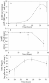
FIG. 1.
FIG. 1.
(A) Growth of P. putida in Leptothrix medium. The cells enter the stationary phase of growth at ∼20 h. (B) Removal of Mn+2(aq) from growth medium occurs after P. putida enters the stationary phase of growth. (C) The production of extracellular polysaccharides (biofilm) during growth. The arrow indicates for each graph where the cells enter the stationary phase of growth. susp., suspension.
FIG. 2.
FIG. 2.
Electron micrographs of P. putida collected during the stationary phase of growth. (A and B) SEM images show the cell oxide aggregates (∼10 by 30 μm) and the associated desiccated biofilm (extracellular polysaccharide material), which is seen as strings or ropes. (C and D) TEM images show the biogenic Mn oxide particles (electron-dense wafers) surrounding the bacterial cell. Scale bar = 2 μm (A), 0.5 μm (B), 0.2 μm (C), or 0.1 μm (D).
FIG. 3.
FIG. 3.
Mn L3edge NEXAFS spectra extracted from three successive stacks, recorded from 620 to 690 eV, on the same spatial region of hydrated acid birnessite. The extent of the chemical changes induced by soft X-rays can be observed in (A) acid birnessite suspended in MQ water and to a much greater extent in (B) acid birnessite suspended in organic growth medium. In each panel the first, second, and third stacks collected are presented from bottom to top.
FIG. 4.
FIG. 4.
(Top) STXM images of the Mn reference materials, MnCl2, Mn2O3 (bixbyite), γ-MnOOH (manganite), and δ-MnIVO2, were collected at the Mn-L3 absorption edge. The spectra were extracted from regions of interest (I), which are encircled by white lines, and normalized by the I0 region, encircled by black lines. (Bottom) The corresponding extracted NEXAFS spectra for the Mn reference materials are presented with the computed atomic absorption used for normalization.
FIG. 5.
FIG. 5.
STXM image sequences or stacks collected during growth of P. putida and Mn oxidation. Samples were collected at t = 16, 24, 34, and 48 h in the 1 mM Mn reaction series (left) and at t = 23, 35, and 47 h in the 0.1 mM Mn reaction series (right). The areas encircled by black lines were used to normalize (I0), and the areas encircled by white lines represent the regions of interest (I).
FIG. 6.
FIG. 6.
Mn L-edge NEXAFS spectra obtained from time-resolved Mn oxidation experiments. The spectra were extracted from the STXM stacks presented in Fig. 5 from areas encircled by the white lines representing I (sample transmitted) and normalized by areas representing I0 (background transmitted) encircled by the black lines. The spectra (bold line) were fit (light line) with linear combinations of Mn(II)Cl2, γ-Mn(III)OOH, and δ-Mn(IV)O2. The numerical results of the spectral fits are presented in Fig. 7.
FIG. 7.
FIG. 7.
Manganese oxidation by P. putida as a function of time with 0.1 and 1 mM initial MnCl2. The relative abundances of Mn(II), Mn(III), and Mn(IV) are calculated by linear fitting of experimental Mn L-edge spectra with reference spectra. The error bars represent uncertainty in the normalization of reference spectra, the choice of reference compounds, and chemical changes due to radiation damage. Values for duplicate sampling areas at time points ≥24 h are displayed and indicate spatial heterogeneity within the samples.

References

    1. Abbate, M., F. M. F. deGroot, J. C. Fuggle, A. Fujimori, Y. Tokura, Y. Fujishima, O. Strebel, M. Domke, G. Kaindl, J. van Elp, B. T. Thole, G. A. Sawatsky, M. Sacchi, and N. Tsuda. 1991. Soft-x-ray absorption studies of the location of extra charges induced by substitution in controlled-valence materials. Phys. Rev. B Condens. Matter 44:419-5422. - PubMed
    1. Auerbach, I., C. Sorensen, H. Hansma, and P. Holden. 2000. Physical morphology and surface properties of unsaturated Pseudomonas putida biofilms. J. Bacteriol. 182:3809-3815. - PMC - PubMed
    1. Banerjee, D., and H. W. Nesbitt. 1999. Oxidation of aqueous Cr(III) at birnessite surfaces: constraints on reaction mechanism. Geochim. Cosmochim. Acta 63:1671-1687.
    1. Banfield, J. F., and R. J. Hamers. 1997. Processes at minerals and surfaces with relevance to microorganisms and prebiotic synthesis, p. 81-122. In J. F. Banfield and K. H. Nealson (ed.), Geomicrobiology: interactions between microbes and minerals. Reviews in Mineralogy, vol. 35. Mineralogical Society of America, Washington, D.C.
    1. Bargar, J. R., B. M. Tebo, U. Bergmann, S. M. Webb, P. Glatzel, V. Q. Chiu, and M. Villalobos. 2005. Biotic and abiotic products of Mn(II) oxidation by spores of the marine Bacillus sp. strain SG-1. Am. Mineral. 90:143-154.

Publication types