. 2005 Jun;32(6):548-52.
doi: 10.1165/rcmb.2005-0059OC. Epub 2005 Mar 3.

Lysozyme secretion by submucosal glands protects the airway from bacterial infection

Affiliations

Lysozyme secretion by submucosal glands protects the airway from bacterial infection

Rana Dajani et al. Am J Respir Cell Mol Biol. 2005 Jun.

Abstract

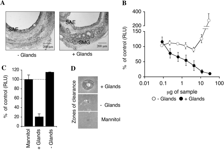
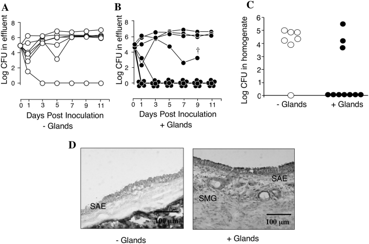
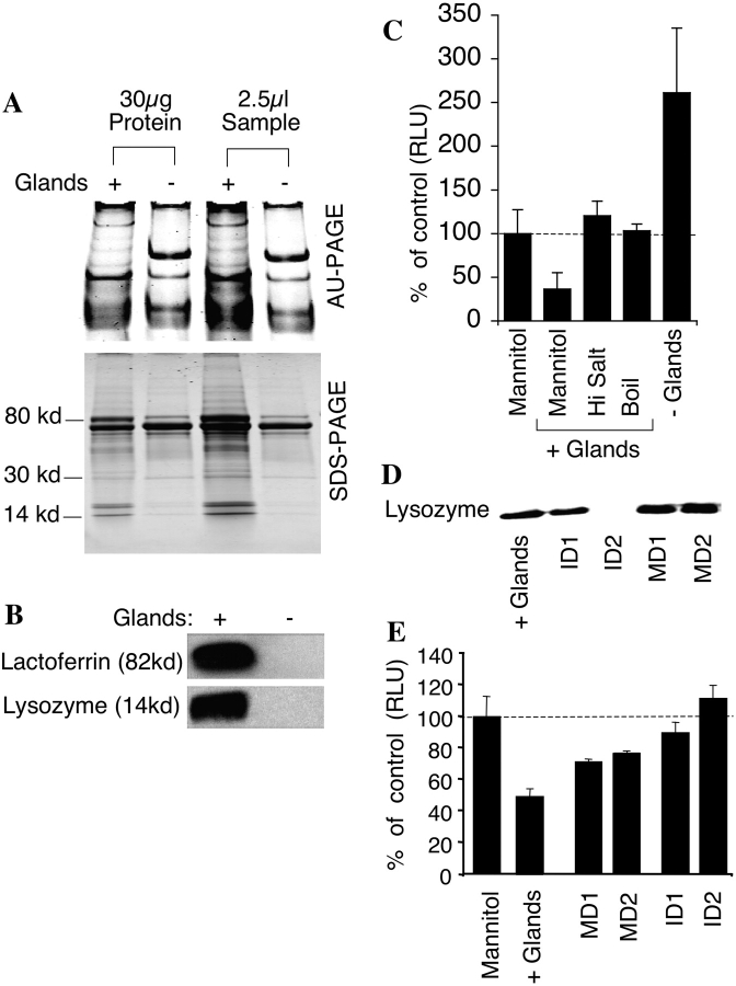
Submucosal glands are abundant (approximately 1 gland/mm2) secretory structures in the tracheobronchial airways of the human lung. Because submucosal glands express antibacterial proteins, it has been proposed that they contribute to lung defense. However, this concept is challenged by the fact that mice do not have submucosal glands in their bronchial airways, yet are quite resistant to bacterial lung infection. The contribution of airway submucosal glands to host defense is also debated as a pathophysiologic component of cystic fibrosis lung disease. Here, we asked whether submucosal glands protect airways against bacterial infection. By comparing tracheal xenograft airways with and without glands, we found that the presence of glands enhanced bacterial killing in vivo and by airway secretions in vitro. Moreover, immunodepletion studies suggested that lysozyme is a major antibacterial component secreted by submucosal glands. These studies provide evidence that submucosal glands are a major source of antibacterials critical for maintaining sterile airways.


Figures

<b>Figure 1.</b>
Figure 1.
Antibacterial activity of ferret xenograft airway secretions. (A) Hematoxylin and eosin (H&E)-stained sections of xenograft trachea with and without glands. SAE, surface airway epithelium; SMG, submucosal glands (bar = 200 μm). (B) The antibacterial activity of equal μg quantities of secretions from tracheal xenografts, with (filled circles) or without (open circles) glands was evaluated using an E. coli bioluminscence assay. (C) The antibacterial activity of 4.3 μg of secretions from xenografts with or without glands against P. aeruginosa was evaluated using a bioluminescence assay. Values in B and C represent the percent of control (5% mannitol) (Mean ± SEM, n = 4 independent samples from 4 different xenografts). (D) The antibacterial activity of 330 μg of tracheal xenograft secretions with and without glands as compared with 5% mannitol (vehicle control) was evaluated using a radial diffusion assay.
<b>Figure 2.</b>
Figure 2.
In vivo survival of bacteria in xenograft airways. (A and B) In vivo survival of bacteria in xenograft airways as measured in the effluent at the indicated time points following in vivo inoculation of 1 × 10⁵ cfu of E. coli. Each xenograft is plotted as a separate line. This xenograft was not able to be perfused after this time point, but upon harvest of the graft was found to be free of bacteria. (C) In vivo survival of E. coli in xenograft tissue homogenates at 14 d after inoculation. Medians are significantly different using a nonparametric Mann Whitney two-tailed assay (P < 0.05), n = 7 independent xenografts without glands (open circles) and n = 10 independent xenografts with glands (filled circles). The same xenografts shown in A and B are shown in C. (D) H&E-stained sections of representative tracheal xenografts without (left panel) with glands (right panel) at 14 d after bacterial inoculation. The xenograft without glands retained bacterial colonization at the time of harvest, whereas the glandular xenograft did not. SAE, surface airway epithelium; SMG, submucosal glands (bar = 100 μm).
<b>Figure 3.</b>
Figure 3.
Analysis of ferret xenograft airway secretions. (A) AU-PAGE and SDS-PAGE of xenograft airway secretions with and without glands. Each lane was loaded with 2.5 μl of secretions or 30 μg of protein. (B) Western blot of lysozyme and lactoferrin from xenograft airway secretions (2.5 μl/lane). (C) The effect of NaCl or boiling on antibacterial activity of xenograft airway secretions. E. coli were incubated with 50 μg of secretions harvested from xenografts with glands in the absence or presence of 125 mM NaCl added to the standard assay buffer. E. coli were also incubated with 50 μg of secretions harvested from xenograft with glands after boiling for 30 min. Values are given as percent of control luminescence (5% mannitol), Mean ± SEM, n = 4. (D) Western blot showing immunodepletion of lysozyme from secretions of xenografts with glands. + Glands, secretions from xenografts with glands; ID1, supernatant following the first immuno-depletion; ID2, supernatant following the second immunodepletion; MD1, supernatant following the first mock immunodepletion; MD2, supernatant following the second mock immunodepletion. All lanes were loaded with 5 μl and are representative of four independent experiments. (E) The effect of immunodepletion of lysozyme from secretions of xenograft with glands on antibacterial activity was measured using the bioluminescent assay on 50 μg of sample. Values represent the percent of control luminescence (5% mannitol), n = 4. Results depict the mean (± SEM) luminescence as compared with control samples.

References

    1. Verkman AS, Song Y, Thiagarajah JR. Role of airway surface liquid and submucosal glands in cystic fibrosis lung disease. Am J Physiol Cell Physiol 2003;284:C2–C15. - PubMed
    1. Welsh MM, Tsui L-C, Boat TF, Beaudet AL, Sly WS, Valle D. The metabolic and molecular bases of inherited disease, 7th ed. New York: McGraw-Hill; 1995.
    1. Tizzano EF, O'Brodovich H, Chitayat D, Benichou JC, Buchwald M. Regional expression of CFTR in developing human respiratory tissues. Am J Respir Cell Mol Biol 1994;10:355–362. - PubMed
    1. Jacquot J, Puchelle E, Hinnrasky J, Fuchey C, Bettinger C, Spilmont C, Bonnet N, Dieterle A, Dreyer D, Pavirani A, et al. Localization of the cystic fibrosis transmembrane conductance regulator in airway secretory glands. Eur Respir J 1993;6:169–176. (see comments). - PubMed
    1. Engelhardt JF, Yankaskas JR, Ernst SA, Yang Y, Marino CR, Boucher RC, Cohn JA, Wilson JM. Submucosal glands are the predominant site of CFTR expression in the human bronchus. Nat Genet 1992;2:240–248. - PubMed

Publication types