Review
. 2005 Mar;62(5):551-77.
doi: 10.1007/s00018-004-4462-3.

G-protein signaling: back to the future

Affiliations
Review

G-protein signaling: back to the future

C R McCudden et al. Cell Mol Life Sci. 2005 Mar.

Abstract

Heterotrimeric G-proteins are intracellular partners of G-protein-coupled receptors (GPCRs). GPCRs act on inactive Galpha.GDP/Gbetagamma heterotrimers to promote GDP release and GTP binding, resulting in liberation of Galpha from Gbetagamma. Galpha.GTP and Gbetagamma target effectors including adenylyl cyclases, phospholipases and ion channels. Signaling is terminated by intrinsic GTPase activity of Galpha and heterotrimer reformation - a cycle accelerated by 'regulators of G-protein signaling' (RGS proteins). Recent studies have identified several unconventional G-protein signaling pathways that diverge from this standard model. Whereas phospholipase C (PLC) beta is activated by Galpha(q) and Gbetagamma, novel PLC isoforms are regulated by both heterotrimeric and Ras-superfamily G-proteins. An Arabidopsis protein has been discovered containing both GPCR and RGS domains within the same protein. Most surprisingly, a receptor-independent Galpha nucleotide cycle that regulates cell division has been delineated in both Caenorhabditis elegans and Drosophila melanogaster. Here, we revisit classical heterotrimeric G-protein signaling and explore these new, non-canonical G-protein signaling pathways.


Figures

Figure 1
Figure 1
Standard model of the GDP/GTP cycle governing activation of heterotrimeric GPCR signaling pathways. In the absence of ligand, the Gα subunit is GDP bound and closely associated with the Gβγ heterodimer. This Gα·GDP/Gβγ heterotrimer interacts with the cytosolic loops of a seven-transmembrane-domain G-protein-coupled receptor (GPCR). Gβγ facilitates the coupling of Gα to receptor and also acts as a guanine nucleotide dissociation inhibitor (GDI) for Gα·GDP, slowing the spontaneous exchange of GDP for GTP. Ligand-bound GPCRs act as guanine nucleotide exchange factors (GEFs) by inducing a conformational change in the Gα subunit, allowing it to exchange GTP for GDP. Gβγ dissociates from Gα·GTP, and both Gα·GTP and Gβγ are competent to signal to their respective effectors. The cycle returns to the basal state when Gα hydrolyzes the gamma-phosphate moiety of GTP, a reaction that is augmented by GTPase-accelerating proteins (GAPs) such as the Regulator of G-protein Signaling (RGS) proteins.
Figure 2
Figure 2
Structural features of heterotrimeric G-protein subunits. (A) The crystal structure of Gα t·GDP·AlF4 (Protein Data Bank identifier: 1TAD) illustrates the Ras-like domain, the all-alpha-helical domain and the bound nucleotide at the interdomain interface. Switch regions I, II and III are shown in blue, GDP in magenta and the phosphate binding loop (P-loop) in yellow. Alpha-helices and beta-sheets are labeled according to traditional designations. (B) Close-up view of the guanine nucleotide binding pocket of a chimeric Gα t/i1 subunit (structural coordinates from PBD ID: 1GOT). Residues that contact the guanine base, ribose sugar, and α and β phosphates are labeled. P-loop residues are shown in yellow and GDP in magenta. (C) The structure of the Gβ 1 γ 1 dimer (PDB ID: 1TBG) shows that Gβ (yellow) forms a seven-bladed propeller consisting of seven WD40 repeats. Gγ (red) forms two alpha helices that bind to the single alpha-helix of Gβ and to several of the WD40 blades. (D) The crystal structure of the heterotrimer (PDB ID: 1GOT) shows that the switch regions of Gα (blue) form part of the interface for interaction with Gβγ.
Figure 3
Figure 3
Schematic of the varied multi-domain architectures of RGS family proteins. RGS subfamily nomenclature follows that first established by Wilkie and Ross [318]. Abbreviations used are Cys (cysteine-rich region), RGS (Regulator of G-protein Signaling domain), DEP (Dishevelled/EGL-10/Pleckstrin homology domain), GGL (Gγ-like domain), PDZ (PSD-95/Dlg/ZO-1 homology domain), PTB (phosphotyrosine binding domain), RBD (Ras binding domain), GoLoco (Gα i/o-Loco interacting motif), βCat (β-catenin binding domain), GSK3β (glycogen synthase kinase-3β binding domain), PP2A (phosphatase PP2A binding domain), DIX (domain present in Dishevelled and Axin), DH (Dbl homology domain), PH (Pleckstrin homology domain), Ser/Thr-kinase (serine-threonine kinase domain).
Figure 4
Figure 4
Domain architecture of mammalian PLC family members. Hallmarks of phosholipase C (PLC) family members are an N-terminal PH domain, which binds Gβγ subunits, and EF, X, Y and C2 motifs forming the catalytic core for phosphoinositide hydrolysis. PLC-γ can be activated by Gα q through a unique C-terminal (CT) domain [319], which also acts as a Gα q GAP [111]. Unique to PLC-γ are two Src-homology-2 (SH2) domains and a Src-homology-3 (SH3) domain that bisect the PH domain. The SH2 domains confer sensitivity to stimulation by PDGF and EGF receptors, whereas the SH3 domain has been shown to act as a GEF for the phosphatidylinositol-3′ kinase (PI3K) enhancer, PIKE [320]. PLC-ɛ interacts with a variety of small GTPases through domains not found in other PLCs. An N-terminal CDC25 (cell division cycle protein 25-like) domain has been shown to promote guanine nucleotide exchange of Ras-family GTPases such as H-Ras and Rap1A, whereas the second Ras-associating (RA) domain (RA2) is reported to bind to H-Ras and Rap in a GTP-dependent fashion; the first RA domain (RA1) displays weak affinity for H-Ras and binds independent of nucleotide state. In addition, RhoA, RhoB and RhoC can activate PLC-ɛ through a unique 60–70-amino acid insert (shaded box) in the Y domain [161]; other Rho family members such as Rac1, Rac2, Rac3 and Cdc42 do not interact with PLC-ɛ.
Figure 5
Figure 5
The 19-amino acid GoLoco motif is found in a diverse set of signaling regulatory proteins. Domain organization of single- and multi-GoLoco motif-containing proteins is illustrated. Abbreviations used are RGS (Regulator of G-protein Signaling domain), RBD (Ras binding domain), GoLoco or GL (Gα i/o-Loco interacting motif), PDZ (PSD-95/Dlg/ZO-1 homology domain), PTB (phosphotyrosine binding domain), RapGAP (Rap-specific GTPase-activating protein domain), GPSM (G-protein-signaling modulator).
Figure 6
Figure 6
Proposed models of AtRGS1 signaling interactions. (A) In the ‘Membrane anchor’ scenario, AtRGS1 acts to recruit (grey arrow) activated AtGPA1 to specific membrane microdomains, allowing localized signaling and deactivation by AtRGS1. (B) In the ‘Spatial focusing’ model, AtRGS1 is a ligand-activated GPCR that coordinately catalyzes both guanine nucleotide exchange on the Arabidopsis heterotrimer (grey arrow) and GTP hydrolysis by the activated Gα subunit (dotted arrow). (C) In the third model, AtRGS1 is a ligand-regulated GAP: i.e. ligand-mediated agonism or inverse agonism regulates deactivation of AtGPA1·GTP via AtRGS1 RGS domain GAP activity.
Figure 7
Figure 7
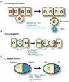
Models of asymmetric cell division in Drosophila and C. elegans. (A) In delaminating neuroblasts, two apical complexes (Bazooka [Baz], atypical protein kinase C [DaPKC] and Par6; Inscuteable [Insc], Partner of Inscuteable [Pins] and Gαi) facilitate the localization of cell-fate determinants to the basal lateral membrane and the orientation of the mitotic spindle. (B) In sensory precursor (SOP) cells, planar polarity is established by counteracting complexes of Baz-DaPKC towards the posterior and Discs Large (Dlg)-Pins-Gαi towards the anterior. (C) In C. elegans one-cell zygotes, PAR-1/-2 proteins enrich GPR-1/2-GOA-1 complex localization towards the posterior, resulting in greater astral microtubule pulling forces on the posterior spindle pole and a resultant smaller P1 daughter cell.
Figure 8
Figure 8
Phenotypes and relative spindle pulling forces of C. elegans embryos in various genetic backgrounds. In wild-type embryos, posterior enrichment of Gα and GPR-1/2 are associated with stronger posterior pulling forces resulting in asymmetric division (light grey, AB daughter cell; dark grey, P1 daughter cell). Loss-of-function mutation or RNAi of either goa-1 or gpa-16 Gα subunit leads to reduction in force magnitude and force asymmetry, but no change in the overall asymmetry of the cell division [294]. Mutation or RNAi of both G-protein subunits, both GoLoco motif proteins gpr-1/2 or the receptor-independent Gα GEF ric-8 causes symmetric division due to loss or mislocalization of pulling force generators. Simultaneous loss of ric-8 and gpb-1 leads to an enhancement of anterior pulling forces indistinguishable from gpb-1 RNAi alone [294]. In contrast, rgs-7 mutants display reduced anterior pulling forces, resulting in exaggerated asymmetry and a smaller P1 cell [305]. In all cases, pulling forces were determined by laser ablation of central mitotic spindles and direct measurement of resultant peak velocities of spindle poles.
Figure 9
Figure 9
Working model of Gα activation during asymmetric cell division of C. elegans embryos. (A) In the wild-type embryo, RIC-8 GEF activity generates Gα·GTP. (It is still unclear whether C. elegans RIC-8 can act directly on Gβγ-complexed Gα·GDP, since rat Ric-8A has been shown, at least in vitro, to act only on free Gα subunits [193]. An alternate possibility is that a distinct pool of free Gα exists or is generated from Gα·GDP/Gβγ heterotrimers by some as-yet unidentified player in this pathway.) The intrinsic GTPase activity of Gα·GTP, possibly accelerated by RGS-7 GAP activity, then generates Gα·GDP that binds the GoLoco motif of GPR-1/2 (the latter protein in complex with its critical co-factor LIN-5; [199]). The GPR-1/2/Gα·GDP complex is presumed to either modulate the (as-yet undefined) astral microtubule force generator or directly generate force (black arrow). (B) In the absence of RIC-8 activity, Gα·GTP levels are reduced, resulting in significantly lower levels of Gα·GDP required to form the GPR-1/2/Gα·GDP complex. This is consistent with the observations of reduced GPR-1/2/Gα·GDP co-immunoprecipitation from ric-8 (md1909+RNAi) C. elegans embryos [294]. (C) Combining the elimination of RIC-8 activity with RNAi-mediated knockdown of gpb-1 (Gβ) is observed to restore the levels of GPR-1/2/Gα·GDP complex [294] and the magnitude of force applied to the spindle poles (fig. 8). Gα·GDP freed from its normal heterotrimeric state has a higher spontaneous nucleotide exchange rate [5] and therefore, in this model, can cycle through the GTP-bound state, GTP hydrolysis and GPR-1/2 interaction (or can directly bind to GPR-1/2; not shown). (D) In this model, loss of RGS-7 GAP activity leads to slower conversion of Gα·GTP to the Gα·GDP form required for the GPR-1/2/Gα·GDP complex. This is consistent with the reduced anterior forces observed in loss-of-function rgs-7 mutants, although it is not known if RGS-7 is restricted in expression to the anterior cortex [305].

Similar articles

Cited by

References

    1. Evanko D. S., Thiyagarajan M. M., Siderovski D. P. and Wedegaertner P. B. (2001) Gbeta gamma isoforms selectively rescue plasma membrane localization and palmitoylation of mutant Galphas and Galphaq. J. Biol. Chem. 276: 23945–23953 - PubMed
    1. Chen C. A. and Manning D. R. (2001) Regulation of G proteins by covalent modification. Oncogene 20: 1643–1652 - PubMed
    1. Robillard L., Ethier N., Lachance M. and Hebert T. E. (2000) Gbetagamma subunit combinations differentially modulate receptor and effector coupling in vivo. Cell Signal. 12: 673–682 - PubMed
    1. Brandt D. R. and Ross E. M. (1985) GTPase activity of the stimulatory GTP-binding regulatory protein of adenylate cyclase, Gs. Accumulation and turnover of enzyme-nucleotide intermediates. J. Biol. Chem. 260: 266–272 - PubMed
    1. Higashijima T., Ferguson K. M., Sternweis P. C., Smigel M. D. and Gilman A. G. (1987) Effects of Mg2+ and the beta gamma-subunit complex on the interactions of guanine nucleotides with G proteins. J. Biol. Chem. 262: 762–766 - PubMed

Publication types

MeSH terms

LinkOut - more resources