. 2004:102:305-20.

Intravitreal human immune globulin in a rabbit model of Staphylococcus aureus toxin-mediated endophthalmitis: a potential adjunct in the treatment of endophthalmitis

Affiliations

Intravitreal human immune globulin in a rabbit model of Staphylococcus aureus toxin-mediated endophthalmitis: a potential adjunct in the treatment of endophthalmitis

Dennis P Han. Trans Am Ophthalmol Soc. 2004.

Abstract

Objectives: To test the feasibility of human immune globulin (IG, Gamimune N, 10%) as a new treatment for endophthalmitis, the ocular tolerance, distribution, and ability of intravitreal IG to attenuate the toxic effects of Staphylococcus aureus culture supernatant were evaluated in a rabbit model.

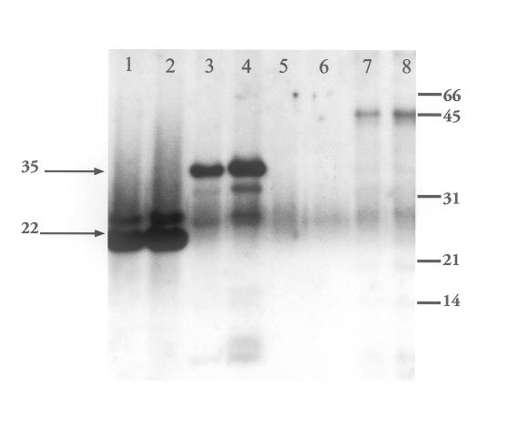
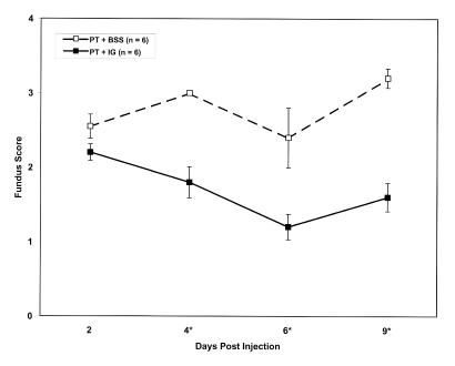
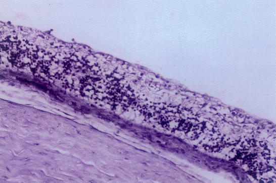
Methods: Effects of intravitreally injected IG were assessed histologically and with Western blot analysis performed 1 to 5 days after injection. IG reactivity to products of S. aureus strain RN4220 was tested by Western blotting, using known toxins (beta hemolysin and toxic shock syndrome toxin-1) and a concentrated culture supernatant containing S. aureus exotoxins (pooled toxin, PT). Endophthalmitis was induced by intravitreal PT injection. For treatment, IG and PT were mixed and injected simultaneously, or IG was injected immediately after, or 6 hours after, PT injection. PT toxicity was graded clinically and histologically over 9 days.

Results: IG persisted intravitreally at least 5 days, inducing no clinical inflammation and minimal mononuclear cell infiltration. In the endophthalmitis model, toxicity from PT was significantly reduced when IG was mixed with PT and injected simultaneously, or when IG was delivered immediately after PT. Only minimal clinically detectable reductions were observed when IG delivery was delayed 6 hours.

Conclusions: Intravitreal IG is well tolerated in the rabbit eye and attenuates the toxicity of culture supernatant containing S. aureus exotoxins. Because toxin elaboration likely occurs gradually in true infection, reduced effects observed with delayed treatment in this toxin-injected model do not preclude clinical application. IG may represent a novel adjunct in endophthalmitis treatment.


Figures

Figure 1
Figure 1
Light micrograph showing the optic nerve head region: mononuclear cells are seen in the prepapillary vitreous following intravitreal injection of human immune globulin (hemtoxylineosin, ×125).
Figure 2
Figure 2
Photomicrograph showing immunolocalization of immunoglobulin molecules following intravitreal injection: intense staining is localized to the inner epithelial layer of the ciliary processes (×125).
Figure 3
Figure 3
Distinct immunoglobulin-specific intracellular staining is seen in the inner nuclear layer of the retina following intravitreal injection of human immune globulin. All the other layers of the neurosensory retina show diffuse staining (×250).
Figure 4
Figure 4
Photomicrograph of the peripapillary retina in the rabbit eye showing diffuse immunoglobulin-specific staining in all the retinal layers. Staining is more intense in the vitreous and in the outer extent of the outer segment layer. The choroid does not show any staining (×250).
Figure 5
Figure 5
Immunoglobulin-specific staining seen in the prepapillary vitreous and around major blood vessels in the optic nerve head, following intravitreal injection of human immune globulin (×125).
Figure 6
Figure 6
Western blot of Staphylococcus aureus culture supernatant (pooled toxin) and purified exotoxins. Nitrocellulose membranes probed and then visualized with ECL detection system. Primary antibody: 1:10,000 dilution of 10% Gamimune; secondary antibody: donkey antihuman. Lanes 1 and 2: toxic shock syndrome toxin-1 (TSST-1) 0.2 and 0.5 μg; lanes 3 and 4: beta hemolysin (BHL) 0.2 and 1.25 μg; lanes 5 and 6: beef heart media; lanes 7 and 8: concentrated culture supernatant 0.5 and 1.0 μg. Molecular weight markers (Bio-Rad) indicated on the right (in kilodaltons) are serum albumin (66), ovalbumin (45), carbonic anhydrase (31), trypsin inhibitor (21), and lysozyme (14). The upper arrow indicates BHL. The lower arrow indicates TSST-1.
Figure 7
Figure 7
Fundus reflex of rabbit eyes 7 days after pooled toxin (PT) injection, without or with Gamimune (IG). Top, PT alone. Bottom, PT, simultaneous IG.
Figure 7
Figure 7
Fundus reflex of rabbit eyes 7 days after pooled toxin (PT) injection, without or with Gamimune (IG). Top, PT alone. Bottom, PT, simultaneous IG.
Figure 8
Figure 8
Time course of fundus scores following injection of pooled toxin (PT), without or with Gamimune (IG). Top, PT and simultaneous injection of balanced salt solution (BSS) or IG. Middle, PT and sequential injection of BSS or IG. Bottom, PT and delayed injection of BSS or IG. Y-axis represents mean fundus score ± standard error. PT and BSS: broken line; PT and IG: solid line. **n = 6 eyes per data point except for postin-jection day 8 in the simultaneous injection group (top) where n = 3 eyes per data point. *Significant difference exists with P value less than .05 calculated from Mann-Whitney test
Figure 8
Figure 8
Time course of fundus scores following injection of pooled toxin (PT), without or with Gamimune (IG). Top, PT and simultaneous injection of balanced salt solution (BSS) or IG. Middle, PT and sequential injection of BSS or IG. Bottom, PT and delayed injection of BSS or IG. Y-axis represents mean fundus score ± standard error. PT and BSS: broken line; PT and IG: solid line. **n = 6 eyes per data point except for postin-jection day 8 in the simultaneous injection group (top) where n = 3 eyes per data point. *Significant difference exists with P value less than .05 calculated from Mann-Whitney test
Figure 9
Figure 9
Histologic score following injection of pooled toxin (PT), without or with Gamimune (IG). Top, PT and simultaneous injection of balanced salt solution (BSS) or IG. Middle, PT and sequential injection of BSS or IG. Bottom, PT and delayed injection of BSS or IG. Y-axis represents mean fundus score ± standard error. PT and BSS: gray bars; PT and IG: black bars. n = 6 eyes per data point. ** All eyes were collected on postinjection day 9 except for 6 eyes (3: PT and BSS; 3: PT and IG) in the simultaneous injection group (top) collected on postinjection day 7. *Significant difference exists with P value less than .05 calculated from Mann-Whitney test.
Figure 9
Figure 9
Histologic score following injection of pooled toxin (PT), without or with Gamimune (IG). Top, PT and simultaneous injection of balanced salt solution (BSS) or IG. Middle, PT and sequential injection of BSS or IG. Bottom, PT and delayed injection of BSS or IG. Y-axis represents mean fundus score ± standard error. PT and BSS: gray bars; PT and IG: black bars. n = 6 eyes per data point. ** All eyes were collected on postinjection day 9 except for 6 eyes (3: PT and BSS; 3: PT and IG) in the simultaneous injection group (top) collected on postinjection day 7. *Significant difference exists with P value less than .05 calculated from Mann-Whitney test.
Figure 10
Figure 10
Histopathology of rabbit eyes 9 days after pooled toxin (PT) injection, without or with Gamimune (IG). Top, PT alone (hematoxylineosin, ×125). Bottom, PT, simultaneous IG (hematoxylineosin, ×82.5).
Figure 10
Figure 10
Histopathology of rabbit eyes 9 days after pooled toxin (PT) injection, without or with Gamimune (IG). Top, PT alone (hematoxylineosin, ×125). Bottom, PT, simultaneous IG (hematoxylineosin, ×82.5).

Similar articles

Cited by

References

    1. Endophthalmitis Vitrectomy Study Group. . Results of the Endophthalmitis Vitrectomy Study: a randomized trial of immediate vitrectomy and intravenous antibiotics for the treatment of postoperative bacterial endophthalmitis. Arch Ophthalmol. 1995;113:1479–1496. - PubMed
    1. Endophthalmitis Vitrectomy Study Group. . Microbiologic factors and visual outcome in the Endophthalmitis Vitrectomy Study. Am J Ophthalmol. 1996;122:830–846. - PubMed
    1. Han DP, Wisniewski SR, Wilson LA, et al. Spectrum and susceptibilities of microbiologic factors isolates in the Endophthalmitis Vitrectomy Study. Am J Ophthalmol. 1996;122:1–17. - PubMed
    1. Kresloff MS, Castellarin AA, Zarbin M. Endophthalmitis. Surv Ophthalmol. 1998;43:193–224. - PubMed
    1. Waheed S, Ritterband DC, Greenfield DS, et al. New patterns of infecting organisms in late bleb-related endophthalmitis: a ten year review. Eye. 1998;12:910–915. - PubMed

Publication types

LinkOut - more resources