. 2004:102:391-422.

Anterior lens capsule management in pediatric cataract surgery

Affiliations

Anterior lens capsule management in pediatric cataract surgery

M Edward Wilson Jr. Trans Am Ophthalmol Soc. 2004.

Abstract

Purpose: To describe and analyze pediatric anterior capsulotomy techniques and make recommendations.

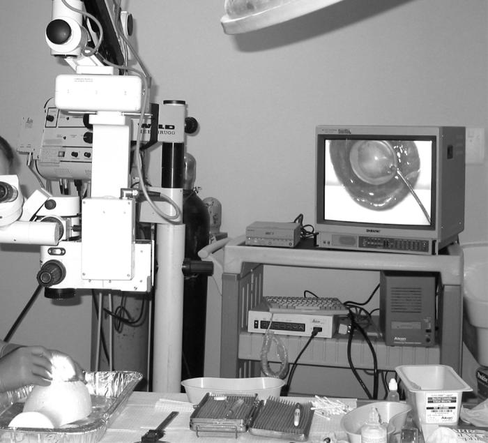
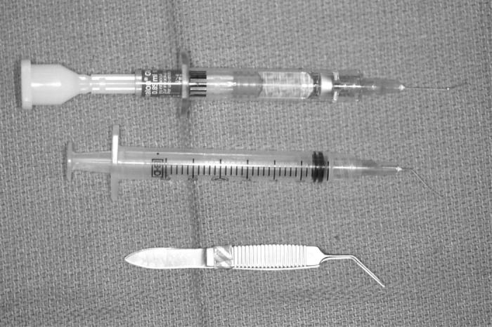
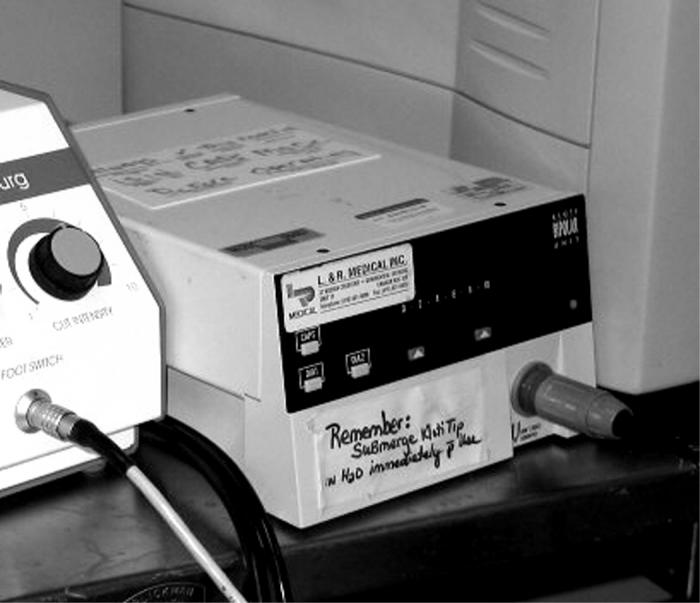
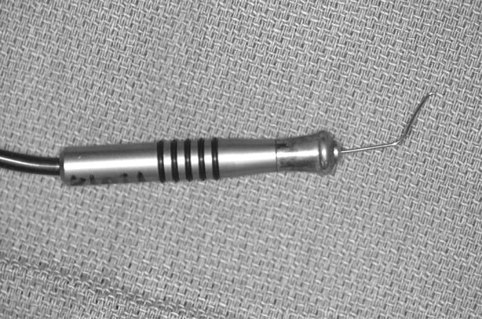
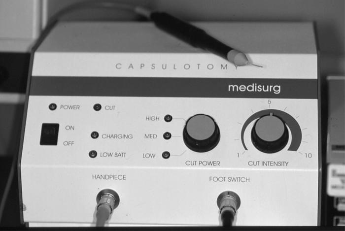
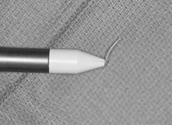
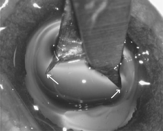
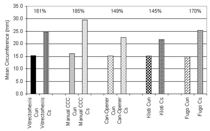
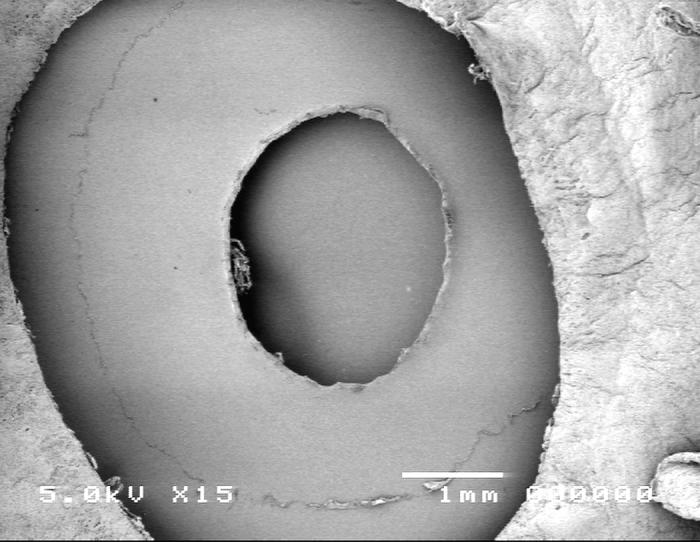
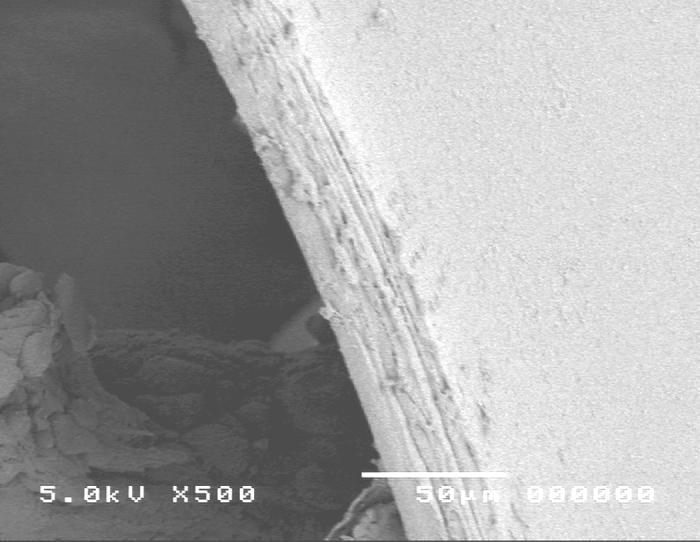
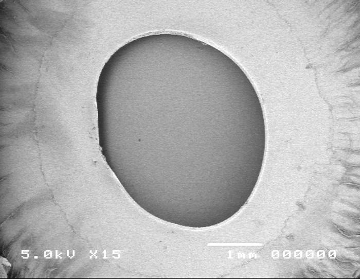
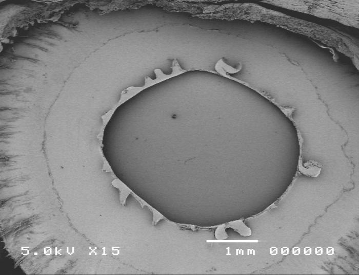
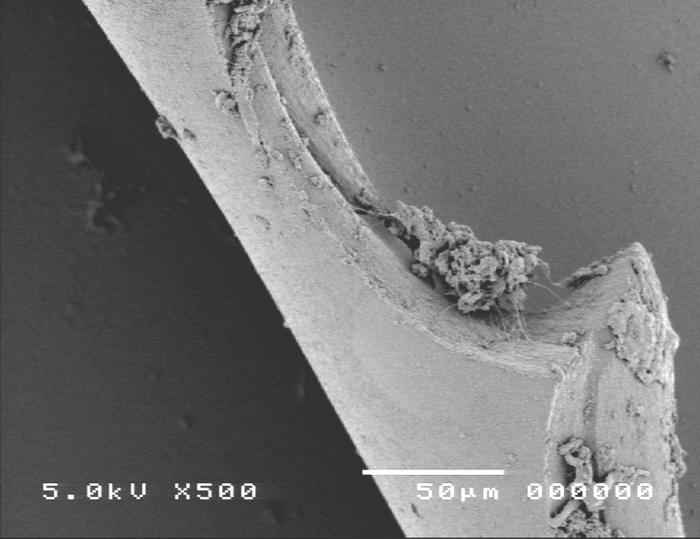
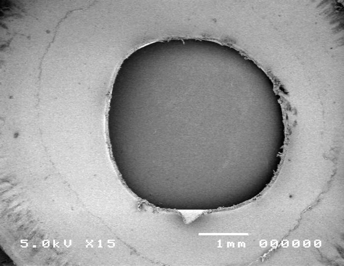
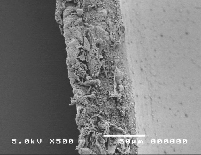
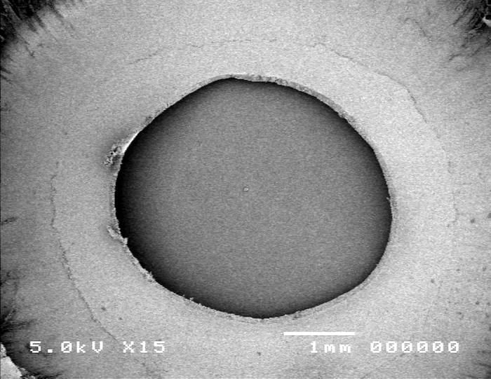
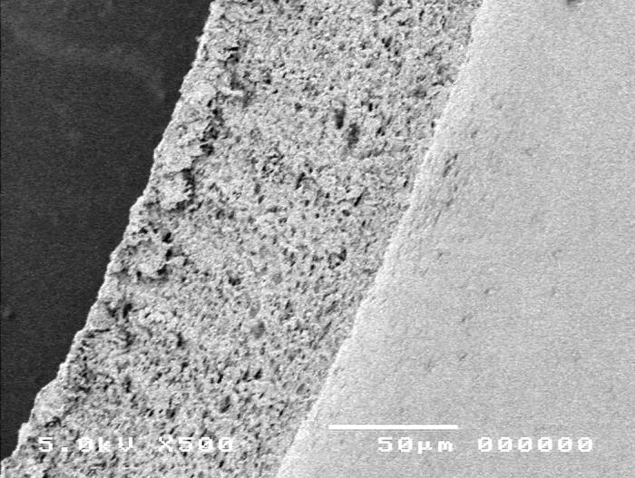
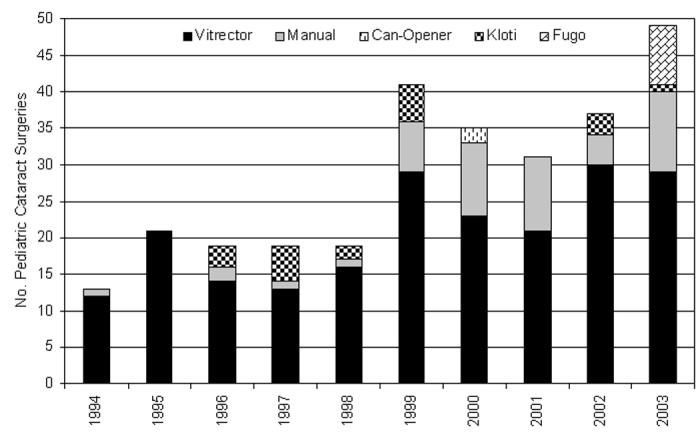
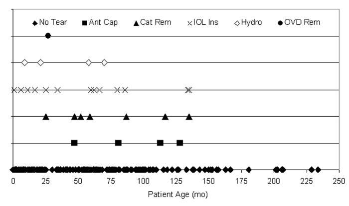
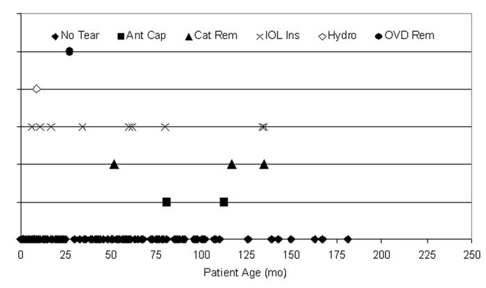
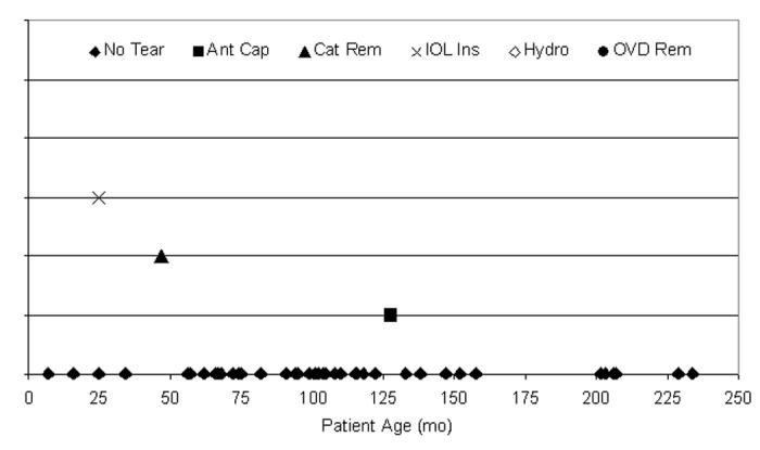
Methods: Five anterior capsulotomy techniques were compared using a porcine model. Extensibility was measured by calculating the mean stretch-to-rupture circumference of each capsulotomy (20 eyes per technique) as a percentage of its circumference at rest. Edge characteristics were reviewed using scanning electron microscopy. A 10-year review of consecutive pediatric cataract surgeries performed by the author focused on the anterior capsulotomy results. A worldwide survey was used to determine current practice patterns.

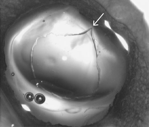
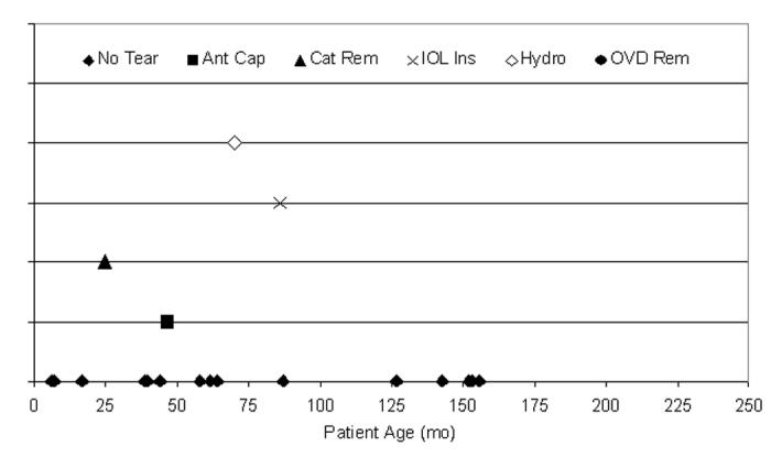
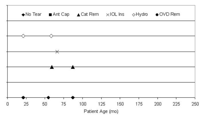
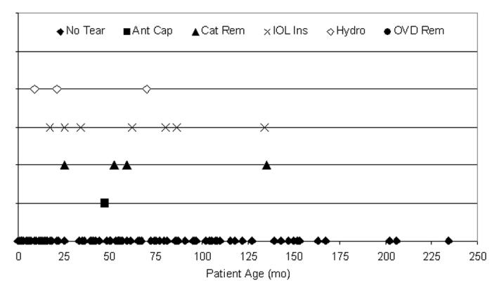
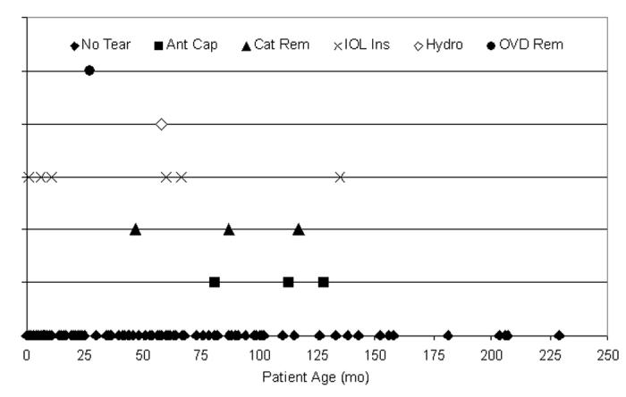
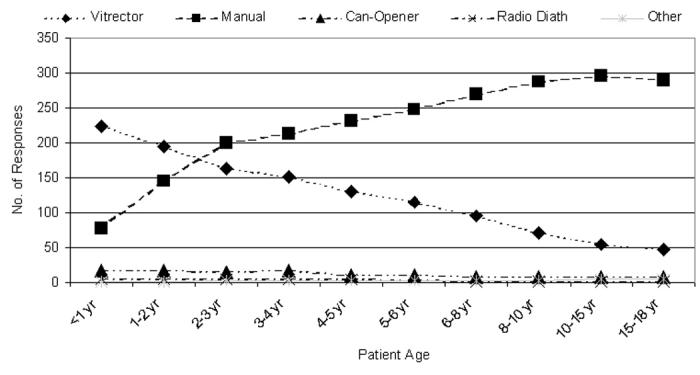
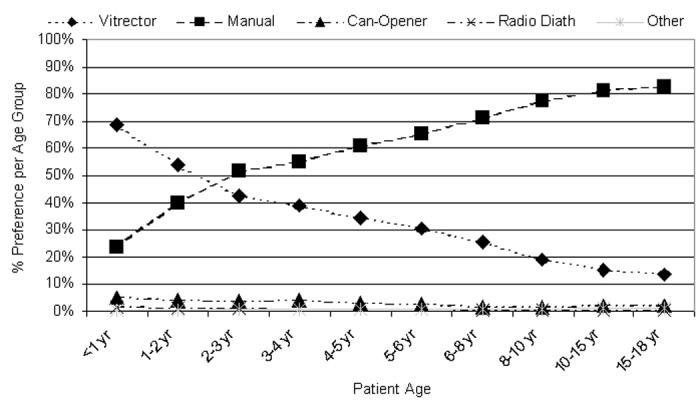
Results: Manual continuous curvilinear capsulorrhexis (CCC) produced the most extensible porcine capsulotomy (185%) with the most regular edge and is preferred by surgeons for patients aged 2 years and older. In the pseudophakic clinical cases reviewed, a radial tear developed in 3 (6.5%) of 46 manual CCC cases. Vitrectorhexis (porcine extensibility, 161%) is preferred by surgeons during the first 2 years of life. A radial tear developed in 16 (7.7%) of 208 vitrectorhexis pseudophakic eyes (29 tears in 284 pseudophakic eyes [10.2%] overall). The Kloti diathermy unit, Fugo plasma blade, and "can-opener" technique produced porcine capsulotomies of 145%, 170%, and 149% extensibility, respectively, and radial tears numbering 4 (21%) of 19, 5 of 8, and 1 of 2, respectively, in the clinical series.

Conclusions: All five capsulotomy techniques are recommendable for children. Only the vitrectorhexis and manual CCC are commonly used today. Vitrectorhexis is well suited for use in infants and young children; manual CCC is best used beyond infancy, and it produces the most stable edge.


Figures

Figure 1
Figure 1
The laboratory surgical suite utilized for the porcine capsulotomy experiments.
Figure 2
Figure 2
Vitrector equipment needed for the vitrectorhexis capsulotomy technique.
Figure 2
Figure 2
Vitrector equipment needed for the vitrectorhexis capsulotomy technique.
Figure 3
Figure 3
Surgical equipment needed for the manual continuous curvilinear capsulorrhexis (CCC) capsulotomy technique.
Figure 4
Figure 4
Surgical equipment needed for the manual multipuncture “can-opener” capsulotomy technique.
Figure 5
Figure 5
Kloti equipment needed for the radiofrequency diathermy capsulotomy technique.
Figure 5
Figure 5
Kloti equipment needed for the radiofrequency diathermy capsulotomy technique.
Figure 6
Figure 6
Fugo equipment needed for the plasma-blade capsulotomy technique.
Figure 6
Figure 6
Fugo equipment needed for the plasma-blade capsulotomy technique.
Figure 7
Figure 7
Calipers were used to measure the unstretched and stretched capsulotomy opening. Left, the caliper tips are placed in the unstretched capsulotomy opening (white arrows indicate the caliper tips). Right, The same stretched capsulotomy opening as appears at left, showing where this opening was torn when stretched (white arrow).
Figure 7
Figure 7
Calipers were used to measure the unstretched and stretched capsulotomy opening. Left, the caliper tips are placed in the unstretched capsulotomy opening (white arrows indicate the caliper tips). Right, The same stretched capsulotomy opening as appears at left, showing where this opening was torn when stretched (white arrow).
Figure 8
Figure 8
Percentage comparison of the mean stretched (Cs) to unstretched (Cus) circumference per capsulotomy technique (n = 20 eyes per technique).
Figure 9
Figure 9
Scanning electron micrograph overview of the vitrector capsulotomy (top) and the cut edge of the anterior lens capsule (bottom).
Figure 9
Figure 9
Scanning electron micrograph overview of the vitrector capsulotomy (top) and the cut edge of the anterior lens capsule (bottom).
Figure 10
Figure 10
Scanning electron micrograph overview of the continuous curvilinear capsulotomy (top) and the cut edge of the anterior lens capsule (bottom).
Figure 10
Figure 10
Scanning electron micrograph overview of the continuous curvilinear capsulotomy (top) and the cut edge of the anterior lens capsule (bottom).
Figure 11
Figure 11
Scanning electron micrograph overview of the multipuncture can-opener capsulotomy (top) and the cut edge of the anterior lens capsule (bottom).
Figure 11
Figure 11
Scanning electron micrograph overview of the multipuncture can-opener capsulotomy (top) and the cut edge of the anterior lens capsule (bottom).
Figure 12
Figure 12
Scanning electron micrograph overview of the Kloti radiofrequency diathermy capsulotomy (top) and the cut edge of the anterior lens capsule (bottom).
Figure 12
Figure 12
Scanning electron micrograph overview of the Kloti radiofrequency diathermy capsulotomy (top) and the cut edge of the anterior lens capsule (bottom).
Figure 13
Figure 13
Scanning electron micrograph overview of the Fugo plasma-blade capsulotomy (top) and the cut edge of the anterior lens capsule (bottom).
Figure 13
Figure 13
Scanning electron micrograph overview of the Fugo plasma-blade capsulotomy (top) and the cut edge of the anterior lens capsule (bottom).
Figure 14
Figure 14
Distribution of 284 pediatric cataract surgeries in which an intraocular lens was inserted (pseudophakic group), including anterior capsulotomy technique used and year performed by one surgeon.
Figure 15
Figure 15
Distribution of tears per one surgeon’s experience. Twenty-nine anterior capsule tears (10.2%) occurred during 284 pediatric cataract surgeries. The incidence of each tear location was not statistically different (P > .05) from any other tear location during the 10-year period. Asterisk indicates 2 data points separated by 8 days; Ant Cap, anterior capsulotomy; Cat Rem, cataract removal; IOL Ins, intraocular lens insertion; Hydro, hydrodissection; OVD Rem, ocular viscosurgical device removal.
Figure 16
Figure 16
Distribution of tears per patient age. Twenty-nine anterior capsule tears (10.2%) occurred during 284 pediatric cataract surgeries, 65.5% (19 of 29) in patients 72 months of age or younger. These tears were not statistically attributable to any single year of life (P > .05). No tears occurred in patients aged 136 months or older. Ant Cap, anterior capsulotomy; Cat Rem, cataract removal; IOL Ins, intraocular lens insertion; Hydro, hydrodissection; OVD Rem, ocular viscosurgical device removal.
Figure 17
Figure 17
Distribution of tears per vitrector anterior capsulotomy technique. Sixteen anterior capsule tears (7.7%) occurred during 208 pediatric cataract surgeries using the vitrector for the anterior capsulotomy; 56.3% of tears (9 of 16) occurred in patients 72 months of age or younger. These tears were not statistically attributable to any single year of life (P > .05).
Figure 18
Figure 18
Distribution of tears per manual continuous curvilinear capsulorrhexis anterior capsulotomy technique. Three anterior capsule tears (6.4%) occurred during 47 pediatric cataract surgeries using manual CCC for the anterior capsulotomy; 66.7% of tears (2 of 3) occurred in patients 72 months of age or younger. These tears were not statistically attributable to any single year of life (P > .05).
Figure 19
Figure 19
Distribution of tears per multipuncture can-opener technique. One anterior capsule tear occurred during two pediatric cataract surgeries using multipuncture can-opener for the anterior capsulotomy; this patient was younger than 72 months of age. This tear could not be statistically analyzed because of the small sample size.
Figure 20
Figure 20
Distribution of tears per Kloti radiofrequency diathermy anterior capsulotomy technique. Four anterior capsule tears (21.1%) occurred during 19 pediatric cataract surgeries using Kloti technique for the anterior capsulotomy; 75.0% of tears (3 of 4) occurred in patients 72 months of age or younger. These tears were not statistically attributable to any single year of life (P > .05).
Figure 21
Figure 21
Distribution of tears per Fugo plasma blade anterior capsulotomy technique. Five anterior capsule tears (62.5%) occurred during eight pediatric cataract surgeries using Fugo technique for the anterior capsulotomy; 80.0% of tears (4 of 5) occurred in patients 72 months of age or younger. These tears could not be statistically analyzed due to the small sample size.
Figure 22
Figure 22
Distribution of tears per eye. Twenty-nine anterior capsule tears (10.2%) occurred during 284 pediatric cataract surgeries: 15 tears (10.1%) in the 148 right eyes (top) and 14 tears (10.3%) in the 136 left eyes (bottom). Eleven of 15 right eye tears (73.3%) and 8 of 14 left eye tears (57.1%) occurred in patients 72 months of age or younger. These tears were not statistically attributable to any single year of life or surgical ocular location (P > .05, respectively).
Figure 22
Figure 22
Distribution of tears per eye. Twenty-nine anterior capsule tears (10.2%) occurred during 284 pediatric cataract surgeries: 15 tears (10.1%) in the 148 right eyes (top) and 14 tears (10.3%) in the 136 left eyes (bottom). Eleven of 15 right eye tears (73.3%) and 8 of 14 left eye tears (57.1%) occurred in patients 72 months of age or younger. These tears were not statistically attributable to any single year of life or surgical ocular location (P > .05, respectively).
Figure 23
Figure 23
Number of questionnaire responses per patient age group per anterior capsulotomy technique (n = 398). Radio Diath, radiofrequency diathermy.
Figure 24
Figure 24
Percentage of preferences per age group per anterior capsulotomy technique (n is available in Tables 6 and 7). Radio Diath, radiofrequency diathermy.

References

    1. Resnikoff S, Kocur I. World sight day: 10 October. World Health Organ 2002. Available at: <http://www.who.int/mediacentre/news/releases/pr79/en/>.
    1. Wilson ME, Pandey SK, Thakur J. Paediatric cataract blindness in the developing world: surgical techniques and intraocular lenses in the new millennium. Br J Ophthalmol. 2003;87:14–19. - PMC - PubMed
    1. Foster A, Gilbert C, Rahi J. Epidemiology of cataract in childhood: a global perspective. J Cataract Refract Surg. 1997;23:601–604. - PubMed
    1. Gilbert C, Foster A. Childhood blindness in the context of VISION 2020—the right to sight. Bull World Health Organ. 2001;79:227–232. - PMC - PubMed
    1. Ahmad K. WHO launches international programme to combat childhood blindness. Lancet. 2002;359:2258. - PubMed

Publication types