Comparative Study
. 2005 Feb;51(2):207-20.
doi: 10.1016/j.jinsphys.2004.10.010.

Comparative analysis of selected genes from Diachasmimorpha longicaudata entomopoxvirus and other poxviruses

Affiliations
Comparative Study

Comparative analysis of selected genes from Diachasmimorpha longicaudata entomopoxvirus and other poxviruses

Y Hashimoto et al. J Insect Physiol. 2005 Feb.

Abstract

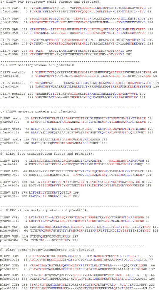
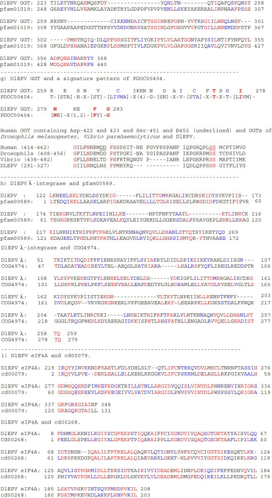
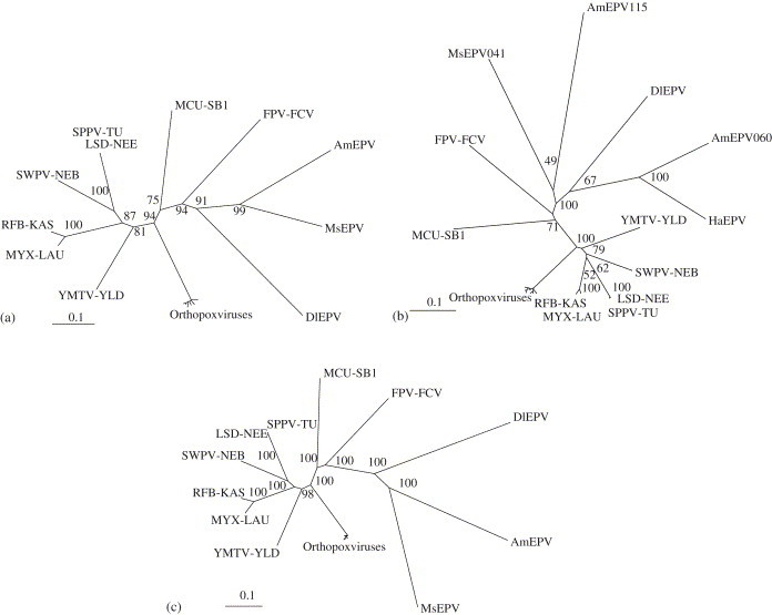
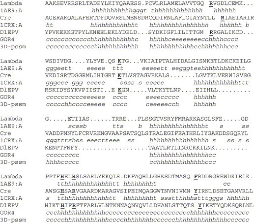
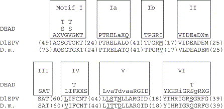
The Diachasmimorpha longicaudata entomopoxvirus (DlEPV) is the first symbiotic EPV described from a parasitic wasp. The DlEPV is introduced into the tephritid fruit fly larval host along with the wasp egg at oviposition. We sequenced a shotgun genomic library of the DlEPV DNA and analyzed and compared the predicted protein sequences of eight ORFs with those of selected poxviruses and other organisms. BlastP searches showed that five of these are homologous to poxvirus putative proteins such as metalloprotease, a putative membrane protein, late transcription factor-3, virion surface protein, and poly (A) polymerase (PAP) regulatory small subunit. Three of these are similar to those of other organisms such as the gamma-glutamyltransferase (GGT) of Arabidopsis thaliana, eukaryotic initiation factor 4A (eIF4A) of Caenorhabditis briggsae and lambda phage integrase (lambda-Int) of Enterococcus faecium. Transcription motifs for early (TGA,A/T,XXXXA) or late (TAAATG, TAAT, or TAAAT) gene expression conserved in poxviruses were identified with those ORFs. Phylogenetic analysis of multiple alignments of five ORFs and 20 poxvirus homologous sequences and of a concatenate of multiple alignments suggested that DlEPV probably diverged from the ancestral node between the fowlpox virus and the genus B, lepidopteran and orthopteran EPVs, to which Amsacta moorei and Melanoplus sanguinipes EPV, respectively, belong. The DlEPV putative GGT, eIF4A, and lambda-Int contained many conserved domains that typified these proteins. These homologues may be involved in either viral pathogenicity or enhancing parasitism via the gamma-glutamyl cycle and compensation of eIF4A levels in the parasitized fly, or via the integration of a portion of the viral genome into the wasp and/or parasitized fly.


Figures

Fig. 1
Fig. 1
Alignment of amino acid sequences of eight DlEPV ORFs with those of the CD or signature sequences of CDs of homologues in the NCBI CD database (http://www.ncbi.nlm.nih.gov;80/Structure/cdd/cdd.shtml). Red=identical; blue=similar; -=gap. (a) DlEPV poly (A) polymerase regulatory subunit (PAP) and the protein family (pfam), pfam01358 (Poly (A) polymerase regulatory subunit) domain. CD-Length=294 residues, 93.9% aligned, E value=1e−47. (b) DlEPV metalloprotease homologue (DlEPV metall) and pfam03410 (Peptidase M44 or Protein G1 that is a glycoprotein expressed by many Poxviridae) domain. CD-Length=590 residues, 28.6% aligned, E value=9e–05. (c) DlEPV membrane protein homologue (DlEPV memb) and pfam02442 (poxvirus unknown orf family) domain. CD-Length=224 residues, 69.6% aligned, E value=2e–17. (d) DlEPV late transcription factor homologue (DlEPV LTF) and pfam04947 (Poxvirus Late Transcription Factor VLTF3 like) domain. CD-Length=218 residues, 91.7% aligned, E value=1e−08. (e) DlEPV virion surface protein homologue (DlEPV VSP) and pfam04584 (Poxvirus A28 family) domain. CD-Length=140 residues, 97.1% aligned, E value=4e−22. (f) DlEPV gamma-glutamyltransferase homologue (DlEPV GGT) and pfam01019 (Gamma-glutamyltranspeptidase) domain. CD-Length=504 residues, 83.9% aligned, E value=7e−42. (g) Upper alignment: DlEPV GGT and a signature pattern of CD of gamma-glutamyltranspeptidase, PDOC00404. AA residues in [ ] represent possible alternative residues. Lower alignment: GGT protein sequences used for alignment are human GGT (AAH25927), Drosophila melanogaster GGT (CAA21412), and Vibrio parahaemolyticus GGT (NP799390). The regions used for alignment are indicated in parenthesis. (h) DlEPV λ-integrase homologue (DlEPV λ) and pfam00589 (Phage integrase) domain. CD-Length=175 residues, 98.3% aligned, E value=1e−09. DlEPV λ-integrase homologue (DlEPV λ) and COG4974 (Site-specific recombinase XerD) domain. CD-Length=300 residues, 77.7% aligned, E value=5e−09. (i) DlEPV eIF4A homologue and cd00079 (Helicase superfamily c-terminal) domain. CD-Length=131 residues, 99.2% aligned, E value=1e−30. DlEPV eIF4A homologue and cd00268 (DEAD-box helicases) domain. CD-Length=203 residues, 100.0% aligned, E value=1e−59.
Fig. 1
Fig. 1
Alignment of amino acid sequences of eight DlEPV ORFs with those of the CD or signature sequences of CDs of homologues in the NCBI CD database (http://www.ncbi.nlm.nih.gov;80/Structure/cdd/cdd.shtml). Red=identical; blue=similar; -=gap. (a) DlEPV poly (A) polymerase regulatory subunit (PAP) and the protein family (pfam), pfam01358 (Poly (A) polymerase regulatory subunit) domain. CD-Length=294 residues, 93.9% aligned, E value=1e−47. (b) DlEPV metalloprotease homologue (DlEPV metall) and pfam03410 (Peptidase M44 or Protein G1 that is a glycoprotein expressed by many Poxviridae) domain. CD-Length=590 residues, 28.6% aligned, E value=9e–05. (c) DlEPV membrane protein homologue (DlEPV memb) and pfam02442 (poxvirus unknown orf family) domain. CD-Length=224 residues, 69.6% aligned, E value=2e–17. (d) DlEPV late transcription factor homologue (DlEPV LTF) and pfam04947 (Poxvirus Late Transcription Factor VLTF3 like) domain. CD-Length=218 residues, 91.7% aligned, E value=1e−08. (e) DlEPV virion surface protein homologue (DlEPV VSP) and pfam04584 (Poxvirus A28 family) domain. CD-Length=140 residues, 97.1% aligned, E value=4e−22. (f) DlEPV gamma-glutamyltransferase homologue (DlEPV GGT) and pfam01019 (Gamma-glutamyltranspeptidase) domain. CD-Length=504 residues, 83.9% aligned, E value=7e−42. (g) Upper alignment: DlEPV GGT and a signature pattern of CD of gamma-glutamyltranspeptidase, PDOC00404. AA residues in [ ] represent possible alternative residues. Lower alignment: GGT protein sequences used for alignment are human GGT (AAH25927), Drosophila melanogaster GGT (CAA21412), and Vibrio parahaemolyticus GGT (NP799390). The regions used for alignment are indicated in parenthesis. (h) DlEPV λ-integrase homologue (DlEPV λ) and pfam00589 (Phage integrase) domain. CD-Length=175 residues, 98.3% aligned, E value=1e−09. DlEPV λ-integrase homologue (DlEPV λ) and COG4974 (Site-specific recombinase XerD) domain. CD-Length=300 residues, 77.7% aligned, E value=5e−09. (i) DlEPV eIF4A homologue and cd00079 (Helicase superfamily c-terminal) domain. CD-Length=131 residues, 99.2% aligned, E value=1e−30. DlEPV eIF4A homologue and cd00268 (DEAD-box helicases) domain. CD-Length=203 residues, 100.0% aligned, E value=1e−59.
Fig. 2
Fig. 2
Unrooted NJ trees of poxvirus virion surface proteins (a), PAP regulatory small subunit (b), and concatenated multiple alignments of virion surface protein, PAP catalytic subunit, metalloprotease, membrane protein and late transcription factor (c). Bootstrap values are shown for each branch. AmEPV=Amsacta moorei entomopoxvirus; DlEPV=Diachasmimorpha longicaudata entomopoxvirus; FPV-FCV= Fowlpox virus; HaEPV=Heliothis armigera entomopoxvirus; LSD-NEE=Lumpy skin disease virus strain Meethling isolate 2490; MCU-SB1=Molluscum contagiosum virus subtype 1; MsEPV=Melanoplus sanguinipes entomopoxvirus; MYX-LAU=Myxoma virus strain Lausanne; RFB-KAS=Rabbit fibroma virus; SPPV-TU =Sheeppox virus TU-V02127; SWPV-NEB=Swinepox virus isolate 17077-99; and YMTV-YLD=Yaba-like disease virus. See Materials and Methods for GenBank accession numbers. The scale bar refers to a phylogenetic distance of 0.1 amino acid substitutions per site.
Fig. 3
Fig. 3
Sequence alignment of the catalytic domain within the putative DlEPV λ-Int (302 aa long) with the bacteriophage homologue (1AE9:A, P03700, 170–357 aa) and Cre recombinase (1CRX:A, P06956, 127–339 aa). Secondary structures of the proteins (1AE9:A and 1CRX:A) were retrieved from the Protein Data Bank (PDB) (http://www.rcsb.org/pdb/). The alignment of λ-Int and Cre recombinase has been reported previously (Nunes-Düby et al., 1998). The key conserved catalytic residues are represented as bold underlined letters (Subramanian et al., 2003). Lambda=Amino acid sequence of the ORF of the bacteriophage λ-integrase. 1AE9:A=C-terminal catalytic domain of bacteriophage λ-integrase. Cre=Amino acid sequence of the ORF of the Cre-recombinase. 1CRX:A=C-terminal catalytic domain of Cre-recombinase. DlEPV=Amino acid sequence of the ORF of DlEPV λ-integrase homologue. GOR4 and 3D-pssm=predicted C-terminal catalytic domain of DlEPV λ-integrase, generated by GOR4 (Garnier et al., 1996) and 3D-pssm (Kelley et al., 2000). Abbreviations for a predicted secondary structure of the query proteins are; b, beta bridge; c, random coil; e, extended strand; g, 310 helix; h, alpha helix; s, bend region; and t, beta turn.
Fig. 4
Fig. 4
Alignments of the conserved motif of DEAD helicases with the eIF4A ORFs of DlEPV (379 aa) and Drosophila melanogaster (D.m.) (403 aa). In the DEAD motif, amino acids conserved at least 80% of the time are shown as capital letters while those conserved 50%-79% of the time are in lower case (Tanner and Linder, 2001). DEAD helicases have 8 motifs (boxed) that are separated into two domains by motif III. Non-conserved amino acid residues between DlEPV and D.m. are underlined. Numbers of amino acid residues between motifs and at both termini are shown in parenthesis.

Similar articles

Cited by

References

    1. Abremski K.E., Hoess R.H. Evidence for a second conserved arginine residue in the integrase family of recombination proteins. Protein Engineering. 1992;5:87–91. - PubMed
    1. Afonso C.L., Tulman E.R., Lu Z., Oma E., Kutish G.F., Rock D.L. The genome of Melanoplus sanguinipes entomopoxvirus. Journal of Virology. 1999;73:533–552. - PMC - PubMed
    1. Afonso C.L., Tulman E.R., Lu Z., Zsak L., Kutish G.F., Rock D.L. The genome of fowlpox virus. Journal of Virology. 2000;74:3815–3831. - PMC - PubMed
    1. Argos P., Landy A., Abremski K., Egan J.B., Haggard-Ljungquist E., Hoess R.H., Kahn M.L., Kalionis B., Narayana S.V., Pierson L.S., III The integrase family of site-specific recombinases: regional similarities and global diversity. The EMBO Journal. 1986;5:433–440. - PMC - PubMed
    1. Azaro M.A., Landy A. The isomeric preference of Holliday junctions influences resolution bias by lambda integrase. The EMBO Journal. 1997;16:3744–3755. - PMC - PubMed

Further reading

    1. Lawrence, P.O., 2000. Parasitism specific proteins and a symbiotic entomopoxvirus from Diachasmimorpha longicaudata, a parasite of tephritid fruit flies. Abstracts of the XXI International Congress of Entomology and XVIII Brazilian Congress of Entomology, Iguassu Falls, Brazil. Abstract #2371, Book II, p. 600.

Publication types