. 2005 Mar 30;334(1):134-43.
doi: 10.1016/j.virol.2005.01.035.

Epitope mapping and biological function analysis of antibodies produced by immunization of mice with an inactivated Chinese isolate of severe acute respiratory syndrome-associated coronavirus (SARS-CoV)

Affiliations

Epitope mapping and biological function analysis of antibodies produced by immunization of mice with an inactivated Chinese isolate of severe acute respiratory syndrome-associated coronavirus (SARS-CoV)

Te-hui W Chou et al. Virology. .

Erratum in

  • Virology. 2005 Jun 20;337(1):204. Mboudoudjeck, Innocent [corrected to Mboudjeka, Innocent]

Abstract

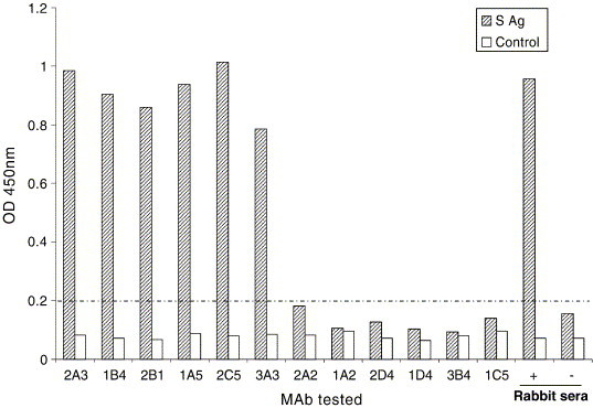
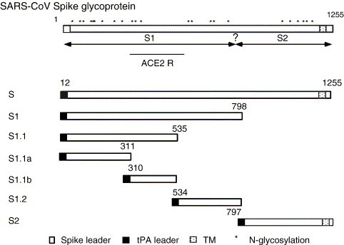
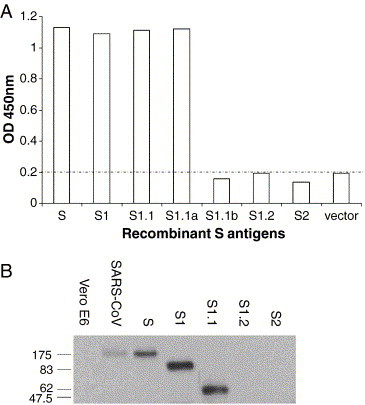
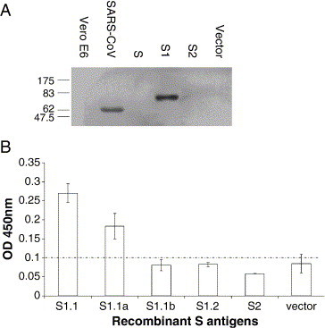
Inactivated severe acute respiratory syndrome-associated coronavirus (SARS-CoV) has been tested as a candidate vaccine against the re-emergence of SARS. In order to understand the efficacy and safety of this approach, it is important to know the antibody specificities generated with inactivated SARS-CoV. In the current study, a panel of twelve monoclonal antibodies (mAbs) was established by immunizing Balb/c mice with the inactivated BJ01 strain of SARS-CoV isolated from the lung tissue of a SARS-infected Chinese patient. These mAbs could recognize SARS-CoV-infected cells by immunofluorescence analysis (IFA). Seven of them were mapped to the specific segments of recombinant spike (S) protein: six on S1 subunit (aa 12-798) and one on S2 subunit (aa 797-1192). High neutralizing titers against SARS-CoV were detected with two mAbs (1A5 and 2C5) targeting at a subdomain of S protein (aa 310-535), consistent with the previous report that this segment of S protein contains the major neutralizing domain. Some of these S-specific mAbs were able to recognize cleaved products of S protein in SARS-CoV-infected Vero E6 cells. None of the remaining five mAbs could recognize either of the recombinant S, N, M, or E antigens by ELISA. This study demonstrated that the inactivated SARS-CoV was able to preserve the immunogenicity of S protein including its major neutralizing domain. The relative ease with which these mAbs were generated against SARS-CoV virions further supports that subunit vaccination with S constructs may also be able to protect animals and perhaps humans. It is somewhat unexpected that no N-specific mAbs were identified albeit anti-N IgG was easily identified in SARS-CoV-infected patients. The availability of this panel of mAbs also provided potentially useful agents with applications in therapy, diagnosis, and basic research of SARS-CoV.


Figures

Fig. 1
Fig. 1
Epitope mapping of mAbs to the S protein by ELISA. MAbs were added at 1:100,000 dilution to each well against either the recombinant full-length S protein (bars in shade) expressed from transiently transfected 293T cells or the vector DNA-transfected 293T cells (bars in blank). “+” and “−” were control sera from New Zealand rabbits immunized with either the full-length S DNA vaccine or the empty vector DNA, respectively. The OD450nm values less than 0.2 are considered non-specific binding to the full-length S antigen in this ELISA assay.
Fig. 2
Fig. 2
Designs of recombinant S segments used in this study. Schematic representation of the entire Spike protein is shown on top, including its natural leader and its transmembrane domain close to the C terminal tail. Predicated N-glycosylation sites are marked by asterisks and the ACE2 receptor binding domain is also noted. DNA plasmids expressing different segments of the S protein were shown in the lower part of the figure with their amino acid residue numbers marked.
Fig. 3
Fig. 3
Epitope mapping of mAb 2A3 using specific recombinant S antigens expressed in 293T transfected cells. (A) Detection of various recombinant S proteins as the coating antigens by ELISA. The OD450nm values in ELISA less than 0.2 are considered non-specific binding to S antigens. (B) Detection of S and its subdomains by Western blot analysis. Samples included uninfected Vero E6 cell lysate (Vero E6), SARS-CoV-infected Vero E6 cell lysate (SARS-CoV), and recombinant S proteins (S, S1, S1.1, S1.2, and S2) as labeled.
Fig. 4
Fig. 4
Detection of SARS-CoV-associated S protein with mAb 3A3 by Western blot analysis. Samples loaded included uninfected Vero E6 lysate and SARS-CoV-infected Vero E6 lysate. Full length S protein (S), the dimmer form of S (di-S), and processed S protein (S′) are indicated.
Fig. 5
Fig. 5
Epitope mapping of mAb 2D4. (A) Western blot analysis of mAb 2D4 with the following samples: uninfected Vero E6 cells, SARS-CoV-infected Vero E6 lysate, recombinant S proteins (S, S1 and S2) expressed from transiently transfected 293T cells, and the control 293T cells transfected with vector DNA. (B) ELISA with mAb 2D4 at 1:500 dilution against different recombinant S antigens (S1.1, S1.1a, S1.1b, S1.2, and S2) expressed from transiently transfected 293T cells and control 293T cells transfected with vector DNA. OD450nm values less than 0.1 are considered non-specific binding in this ELISA assay.
Fig. 6
Fig. 6
Neutralizing activities of mAbs against the Urbani strain of SARS-CoV as measured by neutral red staining in infected Vero E6 cells. Neutralizing activities are plotted as the percent inhibition of viral infection against a particular rabbit serum dilution based on the geometric means from triplet wells. The 50% inhibition (IC50) or 75% inhibition (IC75) levels are marked.

Similar articles

Cited by

References

    1. Arbely E., Khattari Z., Brotons G., Akkawi M., Salditt T., Arkin I.T. A highly unusual palindromic transmembrane helical hairpin formed by SARS coronavirus E protein. J. Mol. Biol. 2004;341(3):769–779. - PMC - PubMed
    1. Berry J.D., Jones S., Drebot M.A., Andonov A., Sabara M., Yuan X.Y., Weingartl H., Fernando L., Marszal P., Gren J., Nicolas B., Andonova M., Ranada F., Gubbins M.J., Ball T.B., Kitching P., Li Y., Kabani A., Plummer F. Development and characterisation of neutralising monoclonal antibody to the SARS-coronavirus. J. Virol. Methods. 2004;120(1):87–96. - PMC - PubMed
    1. Bosch B.J., van der Zee R., de Haan C.A., Rottier P.J. The coronavirus spike protein is a class I virus fusion protein: structural and functional characterization of the fusion core complex. J. Virol. 2003;77(16):8801–8811. - PMC - PubMed
    1. Callow K.A., Parry H.F., Sergeant M., Tyrrell D.A. The time course of the immune response to experimental coronavirus infection of man. Epidemiol. Infect. 1990;105(2):435–446. - PMC - PubMed
    1. Che X.Y., Qiu L.W., Pan Y.X., Xu H., Hao W., Liao Z.Y., Mei Y.B., Zhang L.Y., Wan Z.Y., Yuan G.Y., Huang Z. Rapid and efficient preparation of monoclonal antibodies against SARS-associated coronavirus nucleocapsid protein by immunizing mice. Di Yi Jun Yi Da Xue Xue Bao. 2003;23(7):640–642. - PubMed

Publication types

MeSH terms