

doi: 10.1128/JCM.43.3.1448-1450.2005.
Erroneous reporting of coagulase-negative Staphylococci as Kocuria spp. by the Vitek 2 system
Affiliations
- PMID: 15750130
- PMCID: PMC1081215
- DOI: 10.1128/JCM.43.3.1448-1450.2005
Item in Clipboard
Erroneous reporting of coagulase-negative Staphylococci as Kocuria spp. by the Vitek 2 system
J Clin Microbiol.
2005 Mar.
Abstract
Misidentification of coagulase-negative staphylococci (CoNS) may delay appropriate treatment. We investigated 20 clinical isolates identified as Kocuria spp. by the Vitek 2 system. All were identified as CoNS by 16S rRNA gene sequencing (18 Staphylococcus epidermidis, 1 Staphylococcus haemolyticus). Four Kocuria isolates were shown to be identical to CoNS from the same patient by pulsed-field gel electrophoresis. Isolates identified by Vitek 2 as Kocuria most likely represent misidentified CoNS, and if clinically indicated, should be investigated further by genomic methods.
Figures
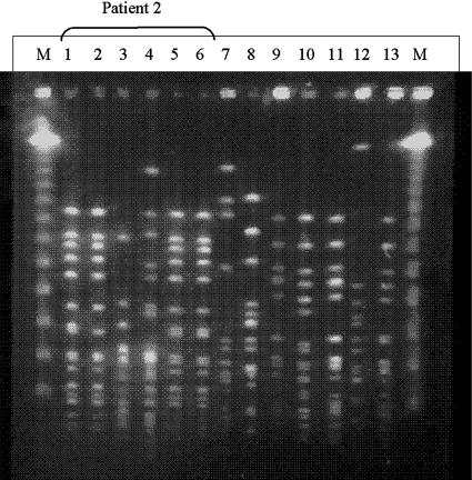
PFGE patterns produced by SmaI macrorestriction from clinical isolates reported as CoNS and Kocuria spp. by Vitek 2. Lanes 1 to 6 contain isolates from patient 2, reported as Kocuria (lanes 1, 2, and 4) and CoNS (lanes 3, 5, and 6). The isolates identified as K. varians from the pacemaker pocket (lane 1) and an electrode (lane 2) were identical to each other, as well as to CoNS isolates from another electrode and blood (lanes 5 and 6, respectively). Another CoNS cultured from blood (lane 3) and an isolate identified as K. varians that was cultured from an electrode (lane 4) were of different clones. Lanes 7 to 13 each contain an isolate identified as Kocuria spp. from a different patient, shown by PFGE to belong to different clones. Lanes M, lambda standard markers. Isolate 7 was identified by 16S rRNA gene sequencing as S. haemolyticus; all other isolates were identified as S. epidermidis.
Comment in
-
Vitek 2 automated identification system and Kocuria kristinae.J Clin Microbiol. 2005 Nov;43(11):5832; author reply 5832. doi: 10.1128/JCM.43.11.5832.2005. J Clin Microbiol. 2005. PMID: 16272536 Free PMC article. No abstract available.
References
-
- Ben-Ami, R., S. Navon-Venezia, D. Schwartz, and Y. Carmeli. 2003. Infection of a ventriculoatrial shunt with phenotypically variable Staphylococcus epidermidis masquerading as polymicrobial bacteremia due to various coagulase-negative staphylococci and Kocuria varians. J. Clin. Microbiol. 41:2444-2447. - PMC - PubMed
MeSH terms
Substances
LinkOut - more resources
Full Text Sources
Miscellaneous
