. 2005 Mar 7;6(1):11.
doi: 10.1186/1471-2121-6-11.

Inclusion of Scar/WAVE3 in a similar complex to Scar/WAVE1 and 2

Affiliations

Inclusion of Scar/WAVE3 in a similar complex to Scar/WAVE1 and 2

Craig F Stovold et al. BMC Cell Biol. .

Abstract

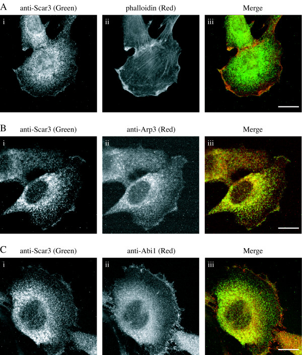
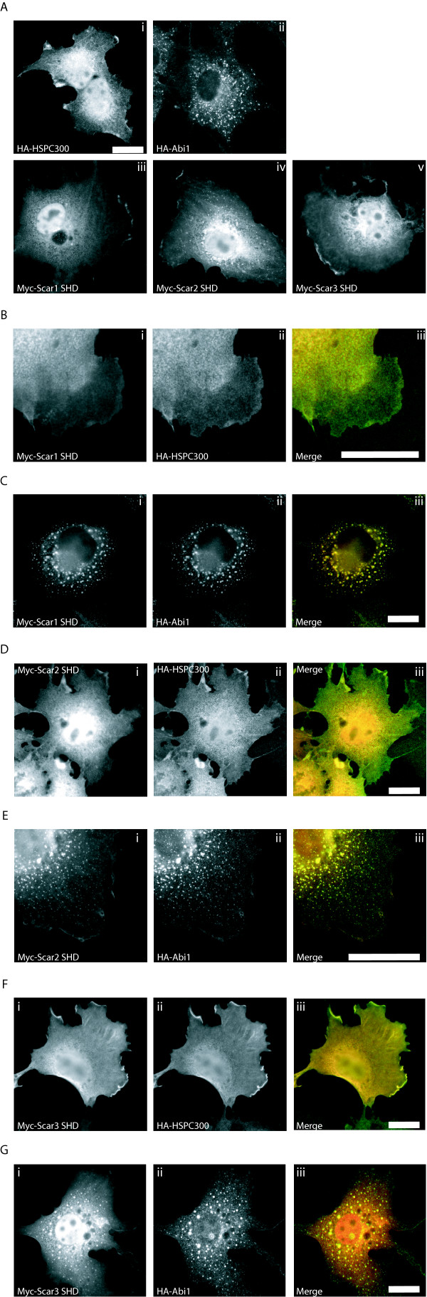
Background: The Scar/WAVE family of proteins mediates signals to actin assembly by direct activation of the Arp2/3 complex. These proteins have been characterised as major regulators of lamellipodia formation downstream of Rac activation and as members of large protein complexes.

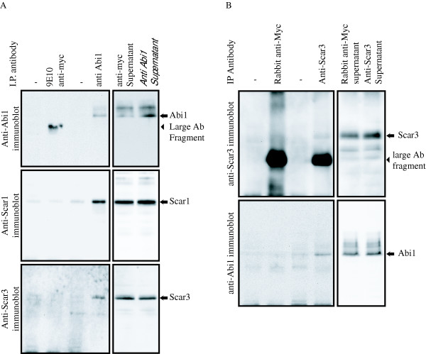
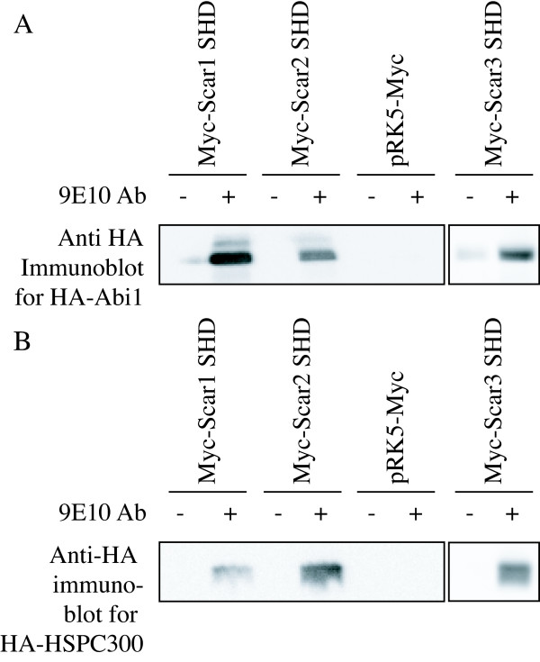
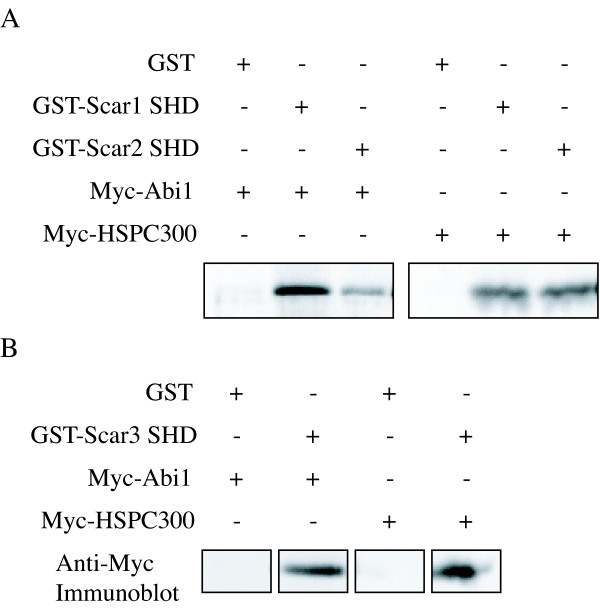
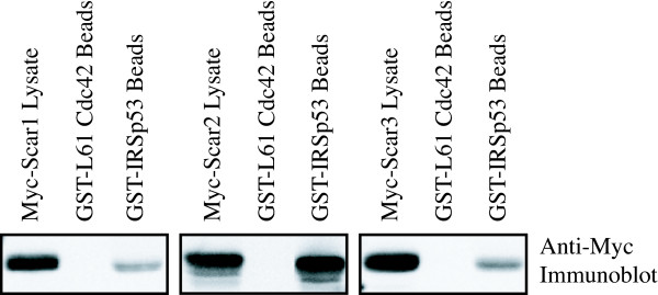
Results: We have investigated the interactions of the three human Scar/WAVE isoforms with several previously described binding partners for Scar/WAVE 1 or 2. We find that all three Scar/WAVE isoforms behave similarly and are likely to participate in the same kinds of protein complexes that regulate actin assembly.

Conclusion: Differences between Scar/WAVE proteins are therefore likely to be at the level of tissue distribution or subtle differences in the affinity for specific binding partners.


Figures

Figure 1
Figure 1
Co-immunoprecipitation of Scar3 and Abi1 from mouse brain extract. Protein G beads were used to precipitate Abi1 from mouse brain extracts in the presence or absence of (A) anti-Abi1 or an unrelated control antibody or (B) anti-Scar3 or an unrelated control antibody. (A) The beads fractions were probed with anti-Abi1, anti-Scar/WAVE1 and anti-Scar/WAVE3. Anti-Abi1 immunoblotting confirmed precipitation of Abi1 only with specific antibody and immunoblotting with anti-Scar/WAVE1 shows association with a known binding partner. Immunoblotting with anti-Scar/WAVE3 revealed Scar/WAVE3 to also be present in Abi1 immunoprecipitates. Anti-Abi1 pulls down both Scar1 and Scar3 together with Abi1. (B) Beads fractions from anti-Scar/WAVE3 immunoprecipitates were probed with both anti-Scar/WAVE3 and anti-Abi1. Both Scar3 and Abi1 are detected in anti-Scar/WAVE3 immunoprecipitates, but not with controls.
Figure 2
Figure 2
Co-immunoprecipitation of Abi1 and HSPC300 with Scar Homology Domain. Cos 7 fibroblasts transiently transfected as indicated. Protein G beads were used to immunoprecipitate Myc-Scar1 SHD, Myc-Scar2 SHD or Myc-Scar3 SHD from lysates in the presence (+) or absence (-) of anti-Myc (9E10) monoclonal antibody. Empty pRK5-Myc vector was used as an additional negative control. (A) HA-Abi1 was detected in the beads plus antibody fractions for Myc-Scar1 SHD, Myc-Scar2 SHD, and Myc-Scar3 SHD, but not in the negative controls. (B) HA-HSPC300 was detected in the beads plus antibody fraction of immunoprecipitations from cells co-transfected only with Myc-Scar1 SHD, Myc-Scar2 SHD, and Myc-Scar3 SHD, but not empty vector controls or in the absence of 9E10 antibody.
Figure 3
Figure 3
Recombinant SHD pulls down Abi1 and HSPC300. Lysate of Cos7 fibroblasts transfected with HA-Abi1 or HSPC300 was incubated as indicated with GST alone, GST-Scar1 SHD (A), GST-Scar2 SHD (A), or GST-Scar3 SHD (B) on glutathione-s-agarose beads. Beads bound fractions were analysed by SDS-PAGE and immunoblotting with a monoclonal anti-Myc (9E10) antibody. HA-Abi1 and HA-HSPC300 were found in pull-downs with all three Scar Homology Domains, but not with GST alone.
Figure 4
Figure 4
IRSp53 interacts with Scar/WAVE1, 2 and 3. Equivalent amounts of GST-IRSp53 or constitutively active GST-L61 Rac were used for pull-down assays from lysates of Cos cells transfected with Myc-Scar1, 2 or 3. Bead fractions and whole cell lysates, indicating loading with each Scar/WAVE isoform, were analysed by SDS-PAGE and immunoblotting with monoclonal (9E10) anti-Myc antibody. None of the Scar/WAVE isoforms bound to active Cdc42, but all were detected in GST-IRSp53 bead fractions.
Figure 5
Figure 5
Cellular localisation of Scar3. C2C12 cells were stained with anti-Scar/WAVE3, anti-Arp3, anti-Abi1 and fluorescently labelled phalloidin to investigate the cellular localisation of endogenous proteins. (A) C2C12 cells stained with (i) anti-Scar/WAVE3 (green), (ii) phalloidin (red) (iii) reveal co-localisation of Scar/WAVE3 with cortical polymerised actin in membrane ruffles. Scar/WAVE3 also colocalises with other areas enriched in polymerised actin, but not stress fibres. (B) Co-staining with (i) anti-Scar/WAVE3 and (ii) anti-Arp3 reveals co-localisation of Scar/WAVE3 (green) with the Arp2/3 complex (red) at protruding areas of the cell membrane. (C) Cells stained with (i) anti-Scar/WAVE3 and (ii) anti-Abi1. (iii) Scar/WAVE3 (green) co-localises with Abi1 (red) in some areas of membrane protrusion. A (i), B (i), and C (i) all show a cytoplasmic and perinuclear and cytoplasmic pool of Scar/WAVE3, with enrichment at areas of lamellipodial protrusion. B (ii) and C (ii) show similar patterns of staining for Arp3 and Abi1. Scale bars are equal to 20 μm in all pictures.
Figure 6
Figure 6
Cellular localization of Abi1, HSPC300, and the Scar homology domain. Transfected Cos cells were stained with a monoclonal anti-Myc antibody and polyclonal anti-HA antibodies to detect over-expressed proteins. (A) Cos7 cells were co-transfected with (i) HA-HSPC300, (ii) HA-Abi1, (iii) Myc-Scar1 SHD, (iv) Myc-Scar2 SHD, or (v) Myc-Scar3 SHD to examine localization of these proteins. HSPC300, and all three SHDs localise in a diffuse cytoplasmic pool with some enrichment at protrusive edges of cells. Abi1 is not detected at the edges of cells, but appears in vesicle-like spots throughout the cytoplasm. (B) Cos7 cells were co-transfected with Myc-Scar1 SHD (i) and HA-HSPC300 (ii) shown as red and green respectively in a merged image (iii). Scar1-SHD and HSPC300 exhibit the same diffuse staining throughout the cytoplasm but also co-localize at protruding edges of cells. (C) Cos7 cells were co-transfected with Myc-Scar1 SHD (i) and HA-Abi1 (ii). (iii) Shows a merge image with myc-Scar1 SHD in red and HA-Abi1 in green. Scar1 SHD and Abi1 both colocalize to punctate spots similar to those seen for Abi1 alone. (D) Cos7 cells co-transfected with Myc-Scar2 SHD (i) and HA-HSPC300 (ii) shown as red and green respectively in a merge image (iii). Both Scar2 SHD and HSPC300 exhibit peri-nuclear staining and localization to protrusive edges of cells. (E) Cos7 cells co-transfected with Myc-Scar2 SHD (i) and HA-Abi1 (ii). (iii) Scar2 SHD (red) and Abi1 (green) show co-localization to cytoplasmic spots and the edges of cells. (F) Cos7 cells co-transfected with (i) Myc-Scar3 SHD (red) and (ii) HA-HSPC300 (green), in a merged image (iii). HSPC300 and Scar3 SHD show diffuse cytoplasmic staining with enrichment at protrusive edges of cells. (G) Cos7 cells co-transfected with (i) Myc-Scar3 SHD and (ii) HA-Abi1. (iii) In a merge image Abi1 (green) and Scar3 SHD (red) co-localize to punctate cytoplasmic spots and to the edges of lamellipodia. Scale bars in all panels are 20 μm.

References

    1. May RC, Machesky LM. Phagocytosis and the actin cytoskeleton. J Cell Sci. 2001;114:1061–1077. - PubMed
    1. May RC, Caron E, Hall A, Machesky LM. Involvement of the Arp2/3 complex in phagocytosis mediated by FcgammaR or CR3. Nat Cell Biol. 2000;2:246–248. doi: 10.1038/35008673. - DOI - PubMed
    1. DeMali KA, Barlow CA, Burridge K. Recruitment of the Arp2/3 complex to vinculin: coupling membrane protrusion to matrix adhesion. J Cell Biol. 2002;159:881–891. doi: 10.1083/jcb.200206043. - DOI - PMC - PubMed
    1. Blanchoin L, Amann KJ, Higgs HN, Marchand JB, Kaiser DA, Pollard TD. Direct observation of dendritic actin filament networks nucleated by Arp2/3 complex and WASP/Scar proteins. Nature. 2000;404:1007–1011. doi: 10.1038/35010008. - DOI - PubMed
    1. Mullins RD, Heuser JA, Pollard TD. The interaction of Arp2/3 complex with actin: nucleation, high affinity pointed end capping, and formation of branching networks of filaments. Proc Natl Acad Sci U S A. 1998;95:6181–6186. doi: 10.1073/pnas.95.11.6181. - DOI - PMC - PubMed

Publication types

LinkOut - more resources