

Association between a variation in the phosphodiesterase 4D gene and bone mineral density
- PMID: 15752431
- PMCID: PMC554993
- DOI: 10.1186/1471-2350-6-9
Association between a variation in the phosphodiesterase 4D gene and bone mineral density
Abstract
Background: Fragility fractures caused by osteoporosis are a major cause of morbidity and mortality in aging populations. Bone mineral density (BMD) is a useful surrogate marker for risk of fracture and is a highly heritable trait. The genetic variants underlying this genetic contribution are largely unknown.
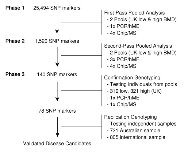
Methods: We performed a large-scale association study investigating more than 25,000 single nucleotide polymorphisms (SNPs) located within 16,000 genes. Allele frequencies were estimated in contrasting DNA pools from white females selected for low (<0.87 g/cm2, n = 319) and high (> 1.11 g/cm2, n = 321) BMD at the lumbar spine. Significant findings were verified in two additional sample collections.
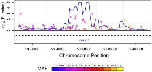
Results: Based on allele frequency differences between DNA pools and subsequent individual genotyping, one of the candidate loci indicated was the phosphodiesterase 4D (PDE4D) gene region on chromosome 5q12. We subsequently tested the marker SNP, rs1498608, in a second sample of 138 white females with low (<0.91 g/cm2) and 138 females with high (>1.04 g/cm2) lumbar spine BMD. Odds ratios were 1.5 (P = 0.035) in the original sample and 2.1 (P = 0.018) in the replication sample. Association fine mapping with 80 SNPs located within 50 kilobases of the marker SNP identified a 20 kilobase region of association containing exon 6 of PDE4D. In a second, family-based replication sample with a preponderance of females with low BMD, rs1498608 showed an opposite relationship with BMD at different sites (p = 0.00044-0.09). We also replicated the previously reported association of the Ser37Ala polymorphism in BMP2, known to interact biologically with PDE4D, with BMD.
Conclusion: This study indicates that variants in the gene encoding PDE4D account for some of the genetic contribution to bone mineral density variation in humans. The contrasting results from different samples indicate that the effect may be context-dependent. PDE4 inhibitors have been shown to increase bone mass in normal and osteopenic mice, but up until now there have been no reports implicating any member of the PDE4 gene family in human osteoporosis.
Figures
Similar articles
-
Evaluation of single nucleotide polymorphisms in the phosphodiesterase 4D gene (PDE4D) and their association with ischaemic stroke in a large German cohort.J Neurol Neurosurg Psychiatry. 2006 Apr;77(4):521-4. doi: 10.1136/jnnp.2005.073577. J Neurol Neurosurg Psychiatry. 2006. PMID: 16543535 Free PMC article.
-
ALOX5AP gene and the PDE4D gene in a central European population of stroke patients.Stroke. 2005 Apr;36(4):731-6. doi: 10.1161/01.STR.0000157587.59821.87. Epub 2005 Feb 24. Stroke. 2005. PMID: 15731479
-
Association between phosphodiesterase 4D gene and ischaemic stroke.J Neurol Neurosurg Psychiatry. 2006 Sep;77(9):1067-9. doi: 10.1136/jnnp.2006.092106. J Neurol Neurosurg Psychiatry. 2006. PMID: 16914755 Free PMC article.
-
PDE4D and stroke: a real advance or a case of the Emperor's new clothes?Stroke. 2006 Aug;37(8):1955-7. doi: 10.1161/01.STR.0000234048.04053.39. Epub 2006 Jul 6. Stroke. 2006. PMID: 16825587 Free PMC article. Review. No abstract available.
-
A close examination of genes within quantitative trait loci of bone mineral density in whole mouse genome.Crit Rev Eukaryot Gene Expr. 2008;18(4):323-43. doi: 10.1615/critreveukargeneexpr.v18.i4.20. Crit Rev Eukaryot Gene Expr. 2008. PMID: 18652562 Review.
Cited by
-
Differences in fat and muscle mass associated with a functional human polymorphism in a post-transcriptional BMP2 gene regulatory element.J Cell Biochem. 2009 Aug 15;107(6):1073-82. doi: 10.1002/jcb.22209. J Cell Biochem. 2009. PMID: 19492344 Free PMC article.
-
Modulation of Bone Morphogenetic Protein (BMP) 2 gene expression by Sp1 transcription factors.Gene. 2007 May 1;392(1-2):221-9. doi: 10.1016/j.gene.2006.12.032. Epub 2007 Jan 20. Gene. 2007. PMID: 17317039 Free PMC article.
-
An autonomous BMP2 regulatory element in mesenchymal cells.J Cell Biochem. 2011 Feb;112(2):666-74. doi: 10.1002/jcb.22975. J Cell Biochem. 2011. PMID: 21268088 Free PMC article.
-
Genetic variation in candidate osteoporosis genes, bone mineral density, and fracture risk: the study of osteoporotic fractures.Calcif Tissue Int. 2008 Sep;83(3):155-66. doi: 10.1007/s00223-008-9165-y. Epub 2008 Sep 12. Calcif Tissue Int. 2008. PMID: 18787887 Free PMC article.
-
MEK Inhibitors Reverse cAMP-Mediated Anxiety in Zebrafish.Chem Biol. 2015 Oct 22;22(10):1335-46. doi: 10.1016/j.chembiol.2015.08.010. Epub 2015 Sep 17. Chem Biol. 2015. PMID: 26388333 Free PMC article.
References
-
- Ray NF, Chan JK, Thamer M, Melton LJ. Medical expenditures for the treatment of osteoporotic fractures in the United States in 1995: report from the National Osteoporosis Foundation. J Bone Miner Res. 1997;12:24–35. - PubMed
-
- Espallargues M, Sampietro-Colom L, Estrada MD, Sola M, del Rio L, Setoain J, Granados A. Identifying bone-mass-related risk factors for fracture to guide bone densitometry measurements: a systematic review of the literature. Osteoporos Int. 2001;12:811–822. doi: 10.1007/s001980170031. - DOI - PubMed
MeSH terms
Substances
LinkOut - more resources
Full Text Sources
Medical
