Comparative Study
. 2005 Mar 14;168(6):941-53.
doi: 10.1083/jcb.200411179. Epub 2005 Mar 7.

ROCK-I regulates closure of the eyelids and ventral body wall by inducing assembly of actomyosin bundles

Affiliations
Comparative Study

ROCK-I regulates closure of the eyelids and ventral body wall by inducing assembly of actomyosin bundles

Yoshihiko Shimizu et al. J Cell Biol. .

Abstract

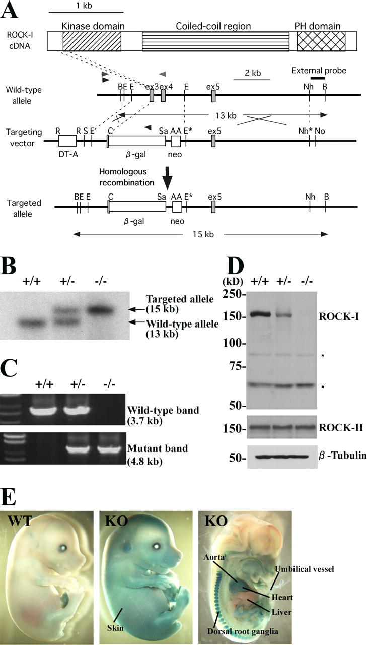
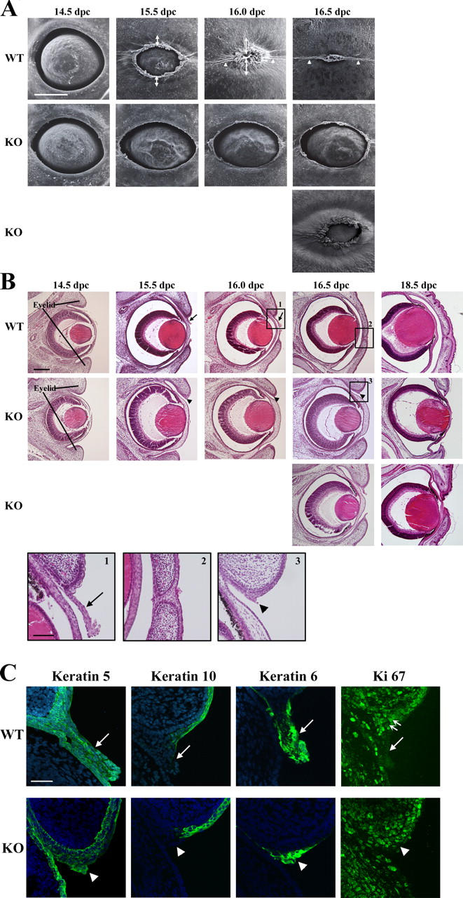
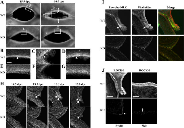
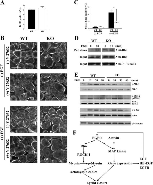
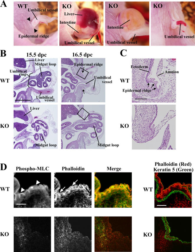
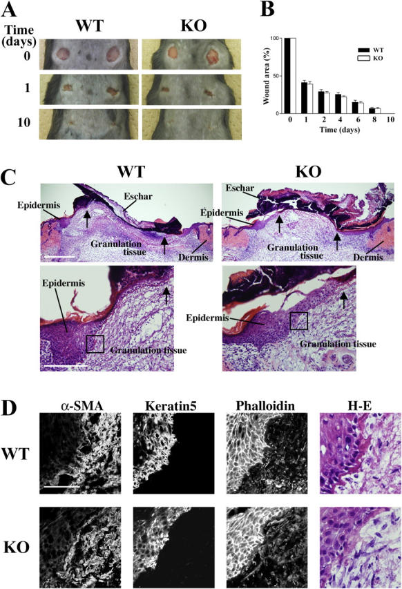
Rho-associated kinase (ROCK) I mediates signaling from Rho to the actin cytoskeleton. To investigate the in vivo functions of ROCK-I, we generated ROCK-I-deficient mice. Loss of ROCK-I resulted in failure of eyelid closure and closure of the ventral body wall, which gave rise to the eyes open at birth and omphalocele phenotypes in neonates. Most ROCK-I(-/-) mice died soon after birth as a result of cannibalization of the omphalocele by the mother. Actin cables that encircle the eye in the epithelial cells of the eyelid were disorganized and accumulation of filamentous actin at the umbilical ring was impaired, with loss of phosphorylation of the myosin regulatory light chain (MLC) at both sites, in ROCK-I(-/-) embryos. Stress fiber formation and MLC phosphorylation induced by EGF were also attenuated in primary keratinocytes from ROCK-I(-/-) mice. These results suggest that ROCK-I regulates closure of the eyelids and ventral body wall through organization of actomyosin bundles.


Figures

Figure 1.
Figure 1.
Generation of ROCK-I–deficient mice. (A) Schematic representations of the domain structure of mouse ROCK-I cDNA, the wild-type ROCK-I allele, the targeting vector, and the targeted allele. Positions of β-galactosidase (β-gal), neomycin resistance (neo), and diphtheria toxin A (DT-A) genes; of restriction sites for BglII (B), EcoRV (E), NheI (Nh), RsrII (R), SmaI (S), ClaI (C), SalI (Sa), AscI (A), and NotI (No); and of exons 3 (ex3), 4, and 5 are shown. The restriction sites indicated by asterisks are lost due to blunt-end ligation. Positions of primers for PCR analysis are indicated by arrowheads. The external probe is a unique 3′ genomic probe that distinguishes the 13-kb wild-type BglII fragment from the 15-kb BglII fragment generated by the targeted allele. (B) Southern blot analysis of genomic DNA obtained from mouse tail. The genotypes of the wild-type, heterozygous, and homozygous knockout mice are shown as +/+, +/−, and −/−, respectively. (C) Genotyping by PCR analysis of genomic DNA from mouse tail. The leftmost lane contains molecular size standards. (D) Immunoblot analysis of whole-brain lysates of adult ROCK-I+/+, ROCK-I+/−, and ROCK-I−/− mice with antibodies specific for either ROCK-I, ROCK-II, or β-tubulin. Positions of molecular mass markers are shown on the left. Asterisks indicate nonspecific bands. (E) X-Gal staining of wild-type (WT) and ROCK-I−/− (KO) embryos at 15.5 dpc (left and middle) or 13.5 dpc (right, sagittal section). X-Gal staining was detected in the skin, heart, aorta, umbilical blood vessels, and dorsal root ganglia of ROCK-I−/− embryos.
Figure 2.
Figure 2.
EOB and omphalocele phenotypes of ROCK-I–deficient mice. (A) Wild-type and ROCK-I−/− embryos at 18.5 dpc. Arrowheads and arrows indicate EOB and omphalocele phenotypes, respectively. Severe, moderate, and mild forms of omphalocele in the mutant embryos are shown from left to right. (B) Eyes of wild-type and ROCK-I−/− mice at 18.5 dpc. The eyes of the ROCK-I−/− embryos are either fully open (middle) or partially open (right; arrow indicates a small hole). (C) Eyes of adult wild-type and ROCK-I−/− mice. (D) Umbilical region of wild-type and ROCK-I−/− neonates. The umbilical ring in the wild-type neonate is closed (arrowhead), whereas that in the ROCK-I−/− neonate remains open (arrow). (E) Visceral organs in the abdominal cavity of the neonates shown in D. The ventral body wall was removed to render the visceral organs visible. Portions of the liver and intestine are absent in the ROCK-I−/− neonate. (F) Gastrointestinal tract from the stomach to the colon of the neonates shown in D and E. A part of the small intestine is absent in the ROCK-I−/− mouse.
Figure 3.
Figure 3.
Impaired eyelid closure in ROCK-I / embryos. (A) Scanning electron micrographs of the eyes of wild-type (top) and ROCK-I−/− (middle and bottom) embryos from 14.5 to 16.5 dpc. Bidirectional arrows indicate extension of the eyelid rim. Arrowheads indicate eyelid fusion in the wild-type embryos. Bar, 500 μm. (B) Hematoxylin-eosin staining of transverse eye sections from wild-type and ROCK-I−/− embryos from 14.5 to 18.5 dpc. Arrows indicate the eyelid epithelial sheet extending from the rim of the eyelid in the wild-type embryos. Eyelid epithelial extension was impaired in mutant embryos, with arrowheads indicating the cell mass at the expected site of sheet formation. Bar, 200 μm. Boxed regions 1–3 are also shown enlarged. Bar, 20 μm. (C) Immunofluorescence staining (green) for keratins 5, 10, and 6 as well as for Ki67 in frozen sections of the eyelids of wild-type or ROCK-I−/− embryos at 16.0 dpc. Nuclei are stained blue. Large arrows indicate the eyelid epithelial sheet; arrowheads indicate the epithelial cell mass. The two small arrows indicate the base of the eyelid epithelial sheet. Bar, 50 μm.
Figure 4.
Figure 4.
ROCK-I–dependent formation of actomyosin cables in the eyelid epithelial sheet. (A) Whole-mount phalloidin staining of the eyelids of wild-type (top) and ROCK-I−/− (bottom) embryos at 15.5 and 16.0 dpc. Bidirectional arrows indicate extension of the eyelid epithelial sheet. Bar, 500 μm. (B–G) Higher magnification views of the boxed regions in A. Arrowheads indicate actin cables in the eyelid epithelial sheet of wild-type embryos. Bidirectional arrows indicate extension of the eyelid epithelial sheet. ROCK-I−/− embryos failed to assemble continuous actin cables. Bar, 50 μm. (H) Phalloidin staining of transverse sections of the eyelid rim of wild-type (top) and ROCK-I−/− (bottom) embryos from 14.5 to 16.0 dpc. A substantial number of actin cables was apparent in a few layers of the eyelid epithelial sheet of wild-type embryos (arrows). Little filamentous-actin accumulation was detected in mutant embryos, with the exception of a few actin bundles in some embryos (arrowhead). Bar, 50 μm. (I) Immunofluorescence staining for phosphorylated MLC (green) and phalloidin staining (red) in sections of the eyelid of wild-type (top) and ROCK-I−/− (bottom) embryos at 16.0 dpc. Phosphorylated MLC was enriched in the eyelid epithelial sheet of wild-type embryos (arrowhead), where it was colocalized with actin cables (arrow). Little phosphorylated MLC staining was detected in ROCK-I−/− embryos. Bar, 50 μm. (J) Immunofluorescence staining for ROCK-I in sections of the eyelid (left) and skin (right) of wild-type (top) and ROCK-I−/− (bottom) embryos at 16.0 dpc. Nonspecific signals are indicated by arrows. Bars, 50 μm.
Figure 5.
Figure 5.
Impairment of EGF-induced formation of actin stress fibers in primary keratinocytes derived from ROCK-I / embryos. (A) Cell proliferation. Primary keratinocytes derived from wild-type or ROCK-I−/− embryos were cultured in the presence of EGF (10 ng/ml) and evaluated for cell proliferation by measurement of incorporation of BrdU. Data are expressed as the percentage of cells that were BrdU positive and are means ± SEM of values from five independent experiments. (B) Phalloidin staining of keratinocytes. Wild-type and ROCK-I−/− keratinocytes were maintained in serum-free medium for 24 h, cultured with or without EGF (10 ng/ml) for 2 h in the absence or presence of 10 μM Y-27632, and then subjected to staining with phalloidin. Bar, 50 μm. (C) Quantitative analysis of EGF-induced stress fiber formation. Cells incubated with or without EGF for 2 h and stained with phalloidin as in B were analyzed for determination of the percentage of cells with stress fibers. A total of 100 cells were examined for each condition. Data are means ± SEM of values from five independent experiments. *, P < 0.05. (D) EGF-induced Rho activation. Wild-type and ROCK-I−/− keratinocytes were cultured in serum-free medium for 24 h, stimulated with EGF (10 ng/ml) for 0 or 10 min, and then subjected to a pull-down assay for the GTP-bound (active) form of Rho. Rho-GTP precipitated from cell lysates was detected by immunoblot analysis with antibodies to Rho (top), and cell lysates (input) were similarly analyzed for the total amounts of Rho (middle) and β-tubulin (bottom). (E) EGF-induced MLC, JNK, and c-Jun phosphorylation. Wild-type and ROCK-I−/− keratinocytes were maintained in serum-free medium for 24 h before stimulation with EGF (10 ng/ml) for the indicated times. Cell lysates were then subjected to immunoblot analysis with antibodies to MLC, to phosphorylated (p-) MLC, to JNK, to phosphorylated JNK, to c-Jun, to phosphorylated c-Jun, and to β-tubulin (control). (F) Proposed role for ROCK-I in mouse eyelid closure. A Rho–ROCK-I–myosin cascade triggered by activation of the EGFR is required for the assembly of the purse stringlike actin cables that contribute to extension of the eyelid epithelial sheet. An autocrine–paracrine pathway mediated by EGFR or activin results in activation of a MAPK cascade and consequent transcription of EGF, heparin-binding EGF (HB-EGF), and EGFR genes.
Figure 6.
Figure 6.
Impaired closure of the umbilical ring in ROCK-I / mice. (A) Omphalocele in ROCK-I−/− embryos. Wild-type and ROCK-I−/− embryos at 18.5 dpc are shown. Severe, moderate, and mild forms of omphalocele are apparent in the mutant mice shown in the center left, center right, and right panels, respectively. Arrowheads indicate the epidermal ridge in the wild-type embryo. (B) Hematoxylin-eosin staining of sagittal sections through the umbilical region of wild-type (top) and ROCK-I−/− (bottom) embryos at 15.5 (left) and 16.5 (right) dpc. Boxes indicate the region of the ectodermal–amnion transition. Arrows indicate the epidermal ridge. Bar, 1 mm. (C) Higher magnification images of the boxed regions in B. Arrows indicate the epidermal ridge in the wild-type embryo. Bar, 200 μm. (D) Staining of the epidermal ridge. Sections of the epidermal ridge of wild-type (top) and ROCK-I−/− (bottom) embryos at 16.5 dpc were stained with an antibody to phosphorylated MLC (green, arrowheads) and phalloidin (red, arrows) in the left panels, and with phalloidin (red) and an antibody to keratin 5 (green) in the right panel. Bars, 50 μm.
Figure 7.
Figure 7.
Wound healing in adult wild-type and ROCK-I / mice. (A) Adult wild-type or ROCK-I−/− mice were shaved on their backs and a 6-mm-diam cylinder of skin (full thickness) was punched out bilaterally. Healing of the wounds was monitored for 10 d. (B) Wound area was expressed as a percentage of the initial value. Quantitative data are means ± SEM (n = 8 wounds for each group). (C) Hematoxylin-eosin staining of the transverse sections of the wound on day 5 from wild-type (left) and ROCK-I−/− mice (right). Arrows indicate the foremost tips of the migrating epithelial sheet. Areas of the migrating epithelial sheet are enlarged in the bottom panels. Bars: (top) 500 μm; (bottom) 200 μm. (D) Staining of the wound edge. Serial sections of the wound from wild-type (top) and ROCK-I−/− (bottom) mice were stained with antibodies to α-SMA, keratin 5, phalloidin, and hematoxylin-eosin (H-E). The fields of view shown correspond to the regions boxed in C. Bar, 50 μm.

References

    1. Amano, M., Y. Fukata, and K. Kaibuchi. 2000. Regulation and functions of Rho-associated kinase. Exp. Cell Res. 261:44–51. - PubMed
    1. Brewer, S., and T. Williams. 2004. Loss of AP-2alpha impacts multiple aspects of ventral body wall development and closure. Dev. Biol. 267:399–417. - PubMed
    1. Brock, J., K. Midwinter, J. Lewis, and P. Martin. 1996. Healing of incisional wounds in the embryonic chick wing bud: characterization of the actin purse-string and demonstration of a requirement for Rho activation. J. Cell Biol. 135:1097–1107. - PMC - PubMed
    1. Eggenschwiler, J., T. Ludwig, P. Fisher, P.A. Leighton, S.M. Tilghman, and A. Efstratiadis. 1997. Mouse mutant embryos overexpressing IGF-II exhibit phenotypic features of the Beckwith-Wiedemann and Simpson-Golabi-Behmel syndromes. Genes Dev. 11:3128–3142. - PMC - PubMed
    1. Etienne-Manneville, S., and A. Hall. 2002. Rho GTPases in cell biology. Nature. 420:629–635. - PubMed

Publication types

MeSH terms