. 2005 Mar 7;201(5):737-46.
doi: 10.1084/jem.20040685.

T cells that cannot respond to TGF-beta escape control by CD4(+)CD25(+) regulatory T cells

Affiliations

T cells that cannot respond to TGF-beta escape control by CD4(+)CD25(+) regulatory T cells

Linda Fahlén et al. J Exp Med. .

Abstract

CD4(+)CD25(+) regulatory T (T reg) cells play a pivotal role in control of the immune response. Transforming growth factor-beta (TGF-beta) has been shown to be required for T reg cell activity; however, precisely how it is involved in the mechanism of suppression is poorly understood. Using the T cell transfer model of colitis, we show here that CD4(+)CD45RB(high) T cells that express a dominant negative TGF-beta receptor type II (dnTbetaRII) and therefore cannot respond to TGF-beta, escape control by T reg cells in vivo. CD4(+)CD25(+) T reg cells from the thymus of dnTbetaRII mice retain the ability to inhibit colitis, suggesting that T cell responsiveness to TGF-beta is not required for the development or peripheral function of thymic-derived T reg cells. In contrast, T reg cell activity among the peripheral dnTbetaRII CD4(+)CD25(+) population is masked by the presence of colitogenic effector cells that cannot be suppressed. Finally, we show that CD4(+)CD25(+) T reg cells develop normally in the absence of TGF-beta1 and retain the ability to suppress colitis in vivo. Importantly, the function of TGF-beta1(-/-) T reg cells was abrogated by anti-TGF-beta monoclonal antibody, indicating that functional TGF-beta can be provided by a non-T reg cell source.


Figures

Figure 1.
Figure 1.
CD4+CD25+ T cells are unable to prevent colitis induced by transfer of dnTβRII CD4+CD45RBhigh cells. RAG-1−/− mice received 2 × 10⁵ CD4+CD45RBhigh cells isolated from either WT or dnTβRII mice. In addition, some mice also received an equivalent number of WT CD4+CD25+ T cells. (A) Data show the mean weight of six animals per group and are representative of three independent experiments. Numbers in parentheses indicate number of animals with clinical signs of disease killed at day 45 and weights for these animals are included in the mean weights. CD4+CD25+ cells confer significant protection from wasting induced by WT CD4+CD45RBhigh cells (P < 0.03) but fail to prevent progressive weight loss induced by dnTβRII CD4+CD45RBhigh cells (NS). (B) Incidence and severity of colitis at the time of sacrifice. (n) Number of mice in each group. Data are pooled from three independent experiments. CD4+CD25+ cells confer significant protection from colitis induced by WT CD4+CD45RBhigh cells (P < 0.001), but fail to prevent colitis induced by dnTβRII CD4+CD45RBhigh cells (NS). (C) Representative photomicrographs of distal colon of RAG-1−/− mice after transfer of CD4+ T cells. Original magnification, 250 (hematoxylin and eosin).
Figure 2.
Figure 2.
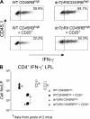
Cytokine production by the progeny of transferred CD4+CD45RBhigh cells isolated from the LP. RAG-1−/− mice received CD4+ T cells as described in Fig. 1. 6–8 wk after transfer, LP cells were isolated and stimulated for 18 h with plate-bound anti-CD3 antibody. (A) Levels of cytokine expression were determined by flow cytometry. Scatter plots are gated on CD4+CD45.1+ lymphocytes to identify progeny of transferred CD4+CD45RBhigh cells and are representative of four to five mice per group. (B) Absolute numbers of cytokine-producing cells were determined by multiplying the frequency of cytokine producing cells after stimulation in vitro by the total number of LP leukocytes. Data are pooled from two independent experiments.
Figure 3.
Figure 3.
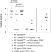
Suppression of colitis by thymic but not splenic dnTβRII CD4+CD25+ cells. RAG-1−/− mice received 2–4 × 10⁵ CD4+CD45RBhigh cells alone or in combination with 1.5–2 × 10⁵ CD4+CD25+ cells isolated from the spleen or thymus of either WT or dnTβRII Tg mice. In addition, some mice received splenic CD4+CD25+ cells alone. Mice were killed 6–8 wk after transfer, and colons were taken for histological analysis. Data show colitis scores for individual mice taken from three independent experiments.
Figure 4.
Figure 4.
CD4+CD25+ cells from dnTβRII mice express normal levels of FoxP3. (A) CD4+CD25+ and CD4+CD25 cells were isolated from spleens of WT (white bars) and dnTβRII (black bars) mice. mRNA was recovered and analyzed for expression of FoxP3 and CD3γ by quantitative real-time PCR. Data show FoxP3 mRNA expression normalized to CD3γ levels as mean ± SEM for triplicate wells. A second experiment gave similar results. (B) Tissue sections from spleens of WT and dnTβRII mice were stained for CD4 (green) and FoxP3 (red). FoxP3+CD4+ cells show red nuclear staining and green surface staining. Frequency of FoxP3+ cells among CD4+ cells: WT 6.4 ± 1.3% and dnTβRII 6.6 ± 1.1%. Numbers represent mean and SEM from three individual mice per group. Original magnification, 200.
Figure 5.
Figure 5.
Frequency of KJ-1.26+ cells is similar among CD4+CD25+ cells from either DO11.10 or DO11.10 TGF-β1−/− mice. Mice analyzed at 6–8 wk of age. CD4 enriched splenocytes from either DO11.10 or DO11.10 TGF-β1−/− mice were analyzed by flow cytometry for the expression of CD4 and CD25. Numbers indicate percentage of CD25+ cells among CD4+ cells. CD4+CD25+ cells were further analyzed for the expression of the clonotypic TCR by mAb KJ-1.26. Numbers indicate percentage KJ-1.26+ cells among CD4+CD25+ cells. Data are representative of six mice per group.
Figure 6.
Figure 6.
CD4+CD25+ T cells from DO11.10.TGF-β1−/− mice retain the ability to suppress colitis. C.B-17 SCID mice received 4 × 10⁵ CD4+CD45RBhigh cells alone or in combination with 105 CD4+CD25+ cells isolated from 6–8-wk-old WT, DO11.10, or DO11.10.TGF-β1−/− mice. Some mice received anti–TGF-β mAb. (A) Incidence and severity of colitis at time of sacrifice. (n) Number of mice in each group. Data are pooled from five independent experiments. CD4+CD25+ cells from WT, DO11.10, and DO11.10 TGF-β1−/− mice confer significant protection from colitis (P < 0.02, P < 0.01, and P < 0.002, respectively). Mice receiving CD4+CD45RBhigh cells together with CD4+CD25+ cells from DO11.10 TGF-β1−/− mice are significantly different from mice that in addition received anti–TGF-β mAb once a week (P < 0.007). (B–D) Representative photomicrographs of colon from C.B-17 SCID mice after transfer of (B) CD4+CD45RBhigh cells, (C and D) CD4+CD45RBhigh, and DO11.10 TGF-β1−/− CD4+CD25+ cells. (D) Mice treated with anti–TGF-β mAb once a week. Original magnification, 200 (hematoxylin and eosin).

Similar articles

Cited by

References

    1. Maloy, K.J., and F. Powrie. 2001. Regulatory T cells in the control of immune pathology. Nat. Immunol. 2:816–822. - PubMed
    1. Shevach, E.M. 2002. CD4+CD25+ suppressor T cells: more questions than answers. Nat. Rev. Immunol. 2:389–400. - PubMed
    1. Takahashi, T., and S. Sakaguchi. 2003. Naturally arising CD25+CD4+ regulatory T cells in maintaining immunologic self-tolerance and preventing autoimmune disease. Curr. Mol. Med. 3:693–706. - PubMed
    1. Sakaguchi, S., N. Sakaguchi, M. Asano, M. Itoh, and M. Toda. 1995. Immunologic self-tolerance maintained by activated T cells expressing IL-2 receptor alpha-chains (CD25). Breakdown of a single mechanism of self-tolerance causes various autoimmune diseases. J. Immunol. 155:1151–1164. - PubMed
    1. Shimizu, J., S. Yamazaki, and S. Sakaguchi. 1999. Induction of tumor immunity by removing CD25+CD4+ T cells: a common basis between tumor immunity and autoimmunity. J. Immunol. 163:5211–5218. - PubMed

Publication types

MeSH terms