. 2005 Mar 15;102(11):4022-6.
doi: 10.1073/pnas.0409219102. Epub 2005 Mar 7.

Differential use of endoplasmic reticulum membrane for phagocytosis in J774 macrophages

Affiliations

Differential use of endoplasmic reticulum membrane for phagocytosis in J774 macrophages

Thalia Becker et al. Proc Natl Acad Sci U S A. .

Abstract

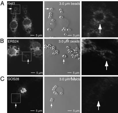
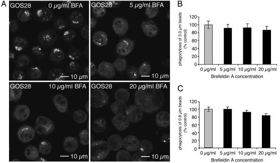
Sustained phagocytosis requires the continuous replacement of cell-surface membrane from intracellular sources. Depending on the nature of the engulfed particles, a variety of endocytic compartments have been demonstrated to contribute membranes needed for the formation of phagosomes. It has recently been reported that the endoplasmic reticulum (ER) can also fuse with the plasma membrane during phagocytosis [Gagnon, E., Duclos, S., Rondeau, C., Chevet, E., Cameron, P. H., Steele-Mortimer, O., Paiement, J., Bergeron, J. J. & Desjardins, M. (2002) Cell 110, 119-131]. However, there is currently no known mechanistic basis for this fusion process to occur. Here we report that direct ER-plasma membrane fusion during phagocytosis requires the ER resident soluble N-ethylmaleimide-sensitive factor attachment protein receptor (SNARE) protein ERS24/Sec22b and that J774-macrophages react toward the challenge of large (3.0-microm) but not small (0.8-microm) particles by triggering this fusion mechanism, allowing them to access the most abundant endogenous membrane source in the cell, the ER.


Figures

Fig. 1.
Fig. 1.
ERS24 is required for phagocytosis of large (3.0-μm) particles. J774 cells were glass-bead-loaded with ERS24 cytoplasmic domain, GST, anti-ERS24 Fab fragments, or random Fab fragments and then challenged with mouse IgG-opsonized latex beads. The measured arbitrary fluorescent intensity of rhodaminedextran reflects the extent of loading. The average number of internalized beads per cell was counted. The extent of bead internalization is expressed as a percentage of the value for unloaded, control cells (set at 100%). (A and B) ERS24 cytoplasmic domain decreases the phagocytosis of 3.0-μm beads (A) but not of 0.8-μm beads (B). (C and D) Anti-ERS24 Fab fragments inhibit phagocytosis of 3.0-μm beads (C) but not of 0.8-μm beads (D) in a dose-dependent manner. (E and F) Inhibition of ERS24 function by ERS24 cytoplasmic domain (E) and anti-ERS24 Fab fragments (F) impairs phagocytosis of aggregates of 0.8-μm beads.
Fig. 4.
Fig. 4.
Phagocytosis of clumped 0.8-μm beads but not of individual 0.8-μm beads induces the mobilization of ER-derived membranes to sites of internalization. J774 cells were pretreated with LY29402 (20 μM) for 30 min before phagocytosis was induced by the addition of mouse IgG-opsonized, 0.8-μm latex beads. (A) Fixed cells were permeabilized and stained with antibodies raised against the SNARE proteins rbet1. rbet1 mobilize to sites of internalization induced by phagocytosis of clumped, 0.8-μm particles (Upper) but not individual 0.8-μm beads (Lower). (B) Pretreatment with two different PI3K inhibitors impairs phagocytosis of mouse IgG-opsonized latex beads [3.0-μm(Upper) and 0.8-μm(Lower)]. J774 cells were pretreated with 0.1 μM wortmannin or 20 μM LY294002 for 30 min and then challenged with phagocytosis in the presence of these inhibitors.
Fig. 3.
Fig. 3.
ER-but not Golgi-derived membranes are recruited for phagocytosis of 3.0-μm beads. J774 cells were pretreated with LY294002 (20 μM) for 30 min before phagocytosis was induced by the addition of mouse IgG-opsonized, 3.0-μm latex beads. (Left) Fixed cells were permeabilized and stained with antibodies raised against rbet1 (A), ERS24 (B), and GOS28 (C). A bead at a site of internalization is marked with a white arrow in each image. (Center) Phase-contrast images of immuno stained regions in Left. A bead at a site of internalization is marked with a white arrow in each image. (Right) Magnified images of respective sites of internalization (arrows). (A) The ER-resident SNARE protein rbet1 mobilizes to the sites of internalization induced by phagocytosis of 3.0-μm beads. (B) The SNARE protein ERS24 is recruited to sites of phagocytosis of 3.0-μm beads. (C) The Golgi-resident SNARE protein GOS28 does not mobilize to sites of internalization.
Fig. 2.
Fig. 2.
Disruption of the Golgi structure by brefeldin A (BFA) does not affect phagocytosis. J774 cells were treated for 30 min with brefeldin A at the concentrations indicated. (A) The distinct Golgi staining of the SNARE protein GOS28 disappears upon treatment with brefeldin A. (B and C) Pretreatment with brefeldin A does not impair phagocytosis of 3.0-μm(B) and 0.8-μm(C) IgG-opsonized latex beads.

References

    1. Cannon, G. J. & Swanson, J. A. (1992) J. Cell Sci. 101, 907–913. - PubMed
    1. Vicker, M. G. (1977) Exp. Cell Res. 109, 127–138. - PubMed
    1. Heine, J. W. & Schnaitman, C. A. (1971) J. Cell Biol. 48, 703–707. - PMC - PubMed
    1. Tardieux, I., Webster, P., Ravesloot, J., Boron, W., Lunn, J. A., Heuser, J. E. & Andrews, N. W. (1992) Cell 71, 1117–1130. - PubMed
    1. Bajno, L., Peng, X. R., Schreiber, A. D., Moore, H. P., Trimble, W. S. & Grinstein, S. (2000) J. Cell Biol. 149, 697–706. - PMC - PubMed

Publication types

MeSH terms