. 2005 Mar 15;102(11):4057-62.
doi: 10.1073/pnas.0501040102. Epub 2005 Mar 7.

Origin and evolution of the chicken leukocyte receptor complex

Affiliations

Origin and evolution of the chicken leukocyte receptor complex

Nikolas Nikolaidis et al. Proc Natl Acad Sci U S A. .

Abstract

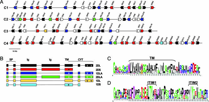
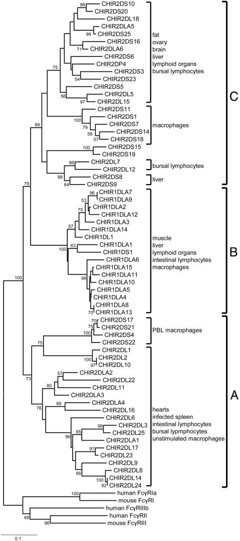
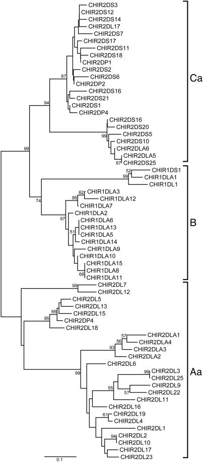
In mammals, the cell surface receptors encoded by the leukocyte receptor complex (LRC) regulate the activity of T lymphocytes and B lymphocytes, as well as that of natural killer cells, and thus provide protection against pathogens and parasites. The chicken genome encodes many Ig-like receptors that are homologous to the LRC receptors. The chicken Ig-like receptor (CHIR) genes are members of a large monophyletic gene family and are organized into genomic clusters, which are in conserved synteny with the mammalian LRC. One-third of CHIR genes encode polypeptide molecules that contain both activating and inhibitory motifs. These genes are present in different phylogenetic groups, suggesting that the primordial CHIR gene could have encoded both types of motifs in a single molecule. In contrast to the mammalian LRC genes, the CHIR genes with similar function (inhibition or activation) are evolutionarily closely related. We propose that, in addition to recombination, single nucleotide substitutions played an important role in the generation of receptors with different functions. Structural models and amino acid analyses of the CHIR proteins reveal the presence of different types of Ig-like domains in the same phylogenetic groups, as well as sharing of conserved residues and conserved changes of residues between different CHIR groups and between CHIRs and LRCs. Our data support the notion that the CHIR gene clusters are regions homologous to the mammalian LRC gene cluster and favor a model of evolution by repeated processes of birth and death (expansion-contraction) of the Ig-like receptor genes.


Figures

Fig. 1.
Fig. 1.
The CHIR gene family. (A) Genomic organization of the CHIR genes. Each arrowhead represents a single gene, and direction of arrowhead shows transcriptional orientation. Genes with similar color have similar structure (see B). Truncated sequences and pseudogenes are shown as white arrowheads. Only sequences homologous to CHIR genes encoding more than three exons are shown. (B) Structure of the CHIR genes. Six types of different CHIR genes were identified. Boxes represent exons, and lines represent introns. Plus sign indicates the presence of the positive residue in the TM region, and Y indicates the ITIM motifs in the CYT region. Coloring of genes corresponds to that in A. (C) Conservation of the TM (in brackets) and CYT regions presented in a logo format. The position of the positive residue is highlighted. (D) Conservation of the CYT region presented in a logo format. The two ITIMs are shown in brackets. Note that alignment gaps were excluded.
Fig. 2.
Fig. 2.
Neighbor-joining tree of the D1 Ig-like domain of the CHIR sequences. The tree was constructed with p distances for 90-aa sites after elimination of alignment gaps. The numbers on the interior branches represent bootstrap values (only values >50 are shown). The sequences of the mammalian Fc receptors were used as outgroups (see also figure 1 of ref. 7). The expression patterns (tissue or cell) of the different CHIRs according to the EST analysis are also shown. The cutoff values for assigning an EST sequence to a particular group of CHIR genes were >90% identity and >90% coverage of the EST sequence. The EST accession numbers are available from M.N. upon request.
Fig. 3.
Fig. 3.
Neighbor-joining tree of the TM-CYT region of the CHIR sequences. The tree was constructed with p distances for 43-aa sites after elimination of alignment gaps.
Fig. 4.
Fig. 4.
Predicted folding of CHIR Ig-like domains. (A) Structural model of the Ig-like domain predicted from the CHIR1DLA10 sequence (in blue) and superimposed on the D1 Ig-like domain of the human KIR2DL1 sequence (in yellow; PDB ID code 1IM9). (B) Structural model of the Ig-like domain predicted from the CHIR2DL15 sequence (in blue) and superimposed on the D1 Ig-like domain of the human LILRB1 sequence (in yellow; PDB ID code 1P7Q). Amino acid residues implicated in ligand binding are shown in red.

References

    1. Lanier, L. L. (2003) Curr. Opin. Immunol. 15, 308–314. - PubMed
    1. Martin, A. M., Kulski, J. K., Witt, C., Pontarotti, P. & Christiansen, F. T. (2002) Trends Immunol. 23, 81–88. - PubMed
    1. Trowsdale, J., Barten, R., Haude, A., Stewart, C. A., Beck, S. & Wilson, M. J. (2001) Immunol. Rev. 181, 20–38. - PubMed
    1. Dennis, G. J., Kubagawa, H. & Cooper, M. D. (2000) Proc. Natl. Acad. Sci. USA 97, 13245–13250. - PMC - PubMed
    1. Volz, A., Wende, H., Laun, K. & Ziegler, A. (2001) Immunol. Rev. 181, 39–51. - PubMed

Publication types

Substances

LinkOut - more resources