. 2005 Mar 15;102(11):4097-102.
doi: 10.1073/pnas.0409832102. Epub 2005 Mar 7.

The protooncogene MYC can break B cell tolerance

Affiliations

The protooncogene MYC can break B cell tolerance

Yosef Refaeli et al. Proc Natl Acad Sci U S A. .

Abstract

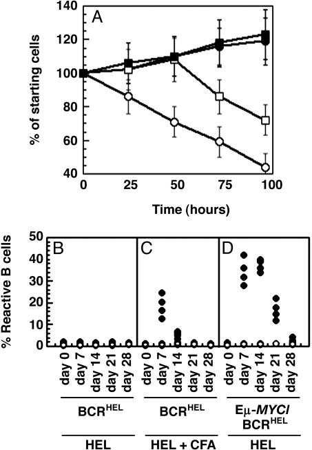
The protooncogene MYC has been implicated in both the proliferation and programmed cell death of lymphoid cells, and in the genesis of lymphoid tumors. Here, we report that overexpression of MYC, as found in many lymphomas, can break immune tolerance. Mice that would otherwise be tolerant to a transgenic autoantigen mounted an immune response to the antigen if MYC was vigorously expressed in the B cell lineage. The responsive B cells converted to an activated phenotype and produced copious amounts of autoantibody that engendered immune complex disease of the kidney. MYC was required to both establish and maintain the breach of tolerance. These effects may be due to the ability of MYC to serve as a surrogate for cytokines. We found that the gene could mimic the effects of cytokines on both B cell proliferation and survival and, indeed, was required for those effects. These findings demonstrate a critical role for MYC in the response of B cells to antigen and expand the potential contributions of MYC to the genesis of lymphomas.


Figures

Fig. 1.
Fig. 1.
The overexpression of MYC restored BCR expression on the surface of anergic B cells. The presentation of BCRHEL on the surface of splenic B cells was determined by flow cytometry. The mice used in AD were age-matched females, bred on a C57BL/6 background (IgMb isotype). Staining for flow cytometry was performed with an antibody to the pan B cell marker B220, and an allotype-specific antibody for the BCRHEL transgene (IgMa). (EH) Mice bearing the MMTV-rtTA and TRE-MYC transgenes were bred on a mixed background. These B cells express both allotypes of IgM, such that IgMa would no longer specifically distinguish the BCRHEL transgene. Staining was with an antibody to B220 and an antibody to HEL (Hy9), to identify antigen bound to the BCRHEL transgene expressed on the surface of the B cells, as described (17). Flow cytometry was performed on spleen cells from a wild-type mouse (C57BL/6) (A), a BCRHEL transgenic mouse (B), a BCRHEL/sHEL mouse (C), an Eμ-MYC/BCRHEL/sHEL mouse (D), wild-type mouse (mixed background) (E), MMTV-rtTA/TRE-MYC/BCRHEL mice that were kept on doxycycline throughout the experiment. (F), MMTV-rtTA/TRE-MYC/BCRHEL/sHEL mice that were kept on doxycycline throughout the experiment (G), or MMTV-rtTA/TRE-MYC/BCRHEL/sHEL mice that had been taken off doxycycline a week before killing (H).
Fig. 2.
Fig. 2.
Accumulation of activated B cells after overexpression of MYC.(A and B) Flow cytometric detection of activated B cells. Analyses were performed on lymph node cells obtained from a wild-type mouse (purple trace), and a 4-week-old Eμ-MYC/BCRHEL/sHEL mouse (green trace). Cells were stained with antibodies to two molecules that are up-regulated after the antigen-dependent activation of B cells, CD69 (A), and B7-2 (CD86) (B). The traces represent the levels of CD69 and B7-2 present on the B220+ fraction of the cells, ascertained by gating on the CyChrome C staining cells by flow cytometry. (C and D) Flow cytometric detection of activated B cells. Analyses were performed on lymph node cells obtained from a wild-type mouse (purple trace), an MMTV-rtTA/TRE-MYC/BCRHEL/sHEL mouse that had been kept on doxycycline throughout (green trace), and an MMTV-rtTA/TRE-MYC/BCRHEL/sHEL mouse that had been taken off doxycycline a week before killing (red trace). Cells were stained with antibodies to two molecules that are up-regulated after the antigen-dependent activation of B cells, CD69 (C), and B7-2 (CD86) (D). The traces represent the levels of CD69 and B7-2 present on the B220+ fraction of the cells, ascertained by gating on the CyChrome C staining cells by flow cytometry. (E and F) Activation of B cells in response to HEL. The percentage of activated B cells in lymph nodes was determined as described for A and B. Each data point in these graphs represents the number of activated B cells detected in the lymph nodes of an individual mouse. Cohorts of four mice were used for each time point. The error bars represent the standard deviations obtained from measurements in triplicate. (E) The accumulation of activated B cells after the induction of MYC overexpression in MMTV-rtTA/TRE-MYC/BCRHEL/sHEL mice. (F) Shows the requirement for MYC in the initiation and maintenance of the accumulation of activated B cells in induced MMTV-rtTA/TRE-MYC/BCRHEL/sHEL mice.
Fig. 3.
Fig. 3.
Accumulation of autoantibodies in serum and immune complexes in the kidneys after the overexpression of MYC.(A and B) Serological evidence of broken tolerance. Sera were obtained from groups of four mice of each of the specified genotypes, and assayed in triplicate by ELISA against HEL (A), or for total serum Ig (B) (see Materials and Methods). The numbered categories represent sera obtained from wild-type mice (bar 1), BCRHEL mice (bar 2), BCRHEL/sHEL mice (bar 3), Eμ-MYC/BCRHEL/sHEL mice before the development of overt tumors (bar 4), MMTV-rtTA/TRE-MYC/BCRHEL/sHEL mice that had been maintained on doxycycline throughout the course of the experiment (bar 5), and MMTV-rtTA/TRE-MYC/BCRHEL/sHEL mice that had been taken off doxycycline 28 days before collection of sera (bar 6). (CF) Immune complex renal disease. Histological examination was performed on kidneys taken from either a wild-type mouse (C) or an Eμ-MYC/BCRHEL/sHEL mouse at 6 weeks of age, before the development of tumor (D). The tissues were sectioned and stained with hematoxylin/eosin, and microscopic images were obtained as described in Materials and Methods. Magnification was ×100. For immununofluorescence, kidneys were obtained from a wild-type mouse (E) or an Eμ-MYC/BCRHEL/sHEL mouse (F). Frozen tissues were sectioned and stained with rhodamine-conjugated antibodies to IgM.
Fig. 4.
Fig. 4.
MYC is an important determinant of B cell proliferation and survival after antigenic stimulation. (A and B) Flow cytometric analysis of cellular proliferation. Splenic B cells were obtained from BCRHEL or Eμ-MYC/BCRHEL mice. The cells were labeled with CFSE and incubated either with antibodies to IgM alone, or a mix of antibodies to IgM and CD40. Flow cytometry was performed after 3 days. (A) The CFSE profile of B cell proliferation for BCRHEL B cells (green trace) and Eμ-MYC/BCRHEL cells by (purple trace). Each peak of the traces represents an additional cell doubling (description of CFSE is contained in Materials and Methods). (B) Overlays the traces obtained for each of the two cell types after stimulation with antibodies to IgM alone (purple trace) or IgM and CD40 (green trace). (C) The effect of MYC on response of B cells to cytokine and antigen. B220+ B cells were activated with antigen and infected with either pMIG (purple traces) or pMIG-Cre (green traces). The cells were then labeled with BODIPY red, incubated overnight with either cytokine or antigen (see Materials and Methods), and analyzed by flow cytometry for GFP expression (to enumerate total cells) and BODIPY red (to detect cellular proliferation). (D and E) Effect of MYC on the survival of B cells. Splenic B cells were activated with antibodies to IgM and CD40 for 4 days. The cells were then washed and incubated in media without added antibodies or growth factors, and assayed for apoptotic cells every 24 h, as described in Materials and Methods.(D) Cells were obtained from MMTV-rtTA/TRE-MYC mice that had either received (clear circles) or not received (filled circles) doxycycline throughout the experiment. (E) Cells were obtained from homozygous c-mycfloxN/floxN mice and then infected with either pMIG (filled circles) or pMIG-Cre (open circles).
Fig. 5.
Fig. 5.
Overexpressed MYC can substitute for B cell cytokines in vitro and T cell help in vivo. (A) Substitution of MYC for cytokine in vitro. Splenic B cells were obtained from IL-4–/– mice and infected with either pMIG or pMIG-MYC. The cells were then incubated in the absence of added cytokine (pMIG, open circles; pMIG-MYC, filled circles), or with a single administration of 1,000 units/ml IL-4 at the beginning of the assay (pMIG, open squares; pMIG-MYC, filled squares). Cells were analyzed as in Fig. 4 D and E.(BD) Substitution of MYC for T cell help in vivo. Spleen and lymph node cells were obtained from either BCRHEL mice (B and C) or Eμ-MYC/BCRHEL mice (D), and transplanted intravenously into wild-type mice. (BD) The mice were then injected with either 100 μg of HEL protein in PBS (filled circles) or PBS alone (open circles). (C) The mice received i.p. injections of 100 μg of HEL protein emulsified in complete Freund's adjuvant (CFA) (filled circles), or PBS emulsified in CFA (open circles). In all instances, spleen and lymph nodes were collected at the indicated times and analyzed by flow cytometry with antibodies specific for B220 and IgMa (same as for Fig. 1 AD).

Similar articles

Cited by

References

    1. Pillai, S. (1999) Immunity 10, 493–502. - PubMed
    1. Goodnow, C. C. (1997) Ann. N.Y. Acad. Sci. 815, 55–66. - PubMed
    1. Nossal, G. J. (1996) J. Exp. Med. 183, 1953–1956. - PMC - PubMed
    1. Adelstein, S., Pritchard-Briscoe, H., Anderson, T. A., Crosbie, J., Gammon, G., Loblay, R. H., Basten, A. & Goodnow, C. C. (1991) Science 251, 1223–1225. - PubMed
    1. Brink, R., Goodnow, C. C., Crosbie, J., Adams, E., Eris, J., Mason, D. Y., Hartley. S. B. & Basten, A. (1992). J. Exp. Med. 176, 991–1005. - PMC - PubMed

Publication types

MeSH terms

LinkOut - more resources