Clinical Trial
. 2005 Apr;54(4):469-78.
doi: 10.1136/gut.2004.041962.

Homeostatic regulation of zinc transporters in the human small intestine by dietary zinc supplementation

Affiliations
Clinical Trial

Homeostatic regulation of zinc transporters in the human small intestine by dietary zinc supplementation

R A Cragg et al. Gut. 2005 Apr.

Abstract

Background: The role of intestinal transporter regulation in optimising nutrient absorption has been studied extensively in rodent and cell line models but not in human subjects.

Aims: The aim of the present study was to investigate the response in vivo of zinc transporters in the human enterocyte to dietary zinc supplementation.

Subjects: Eighteen patients who had previously undergone ileostomy, all free of any symptoms of inflammatory bowel disease.

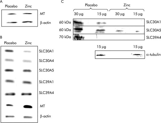
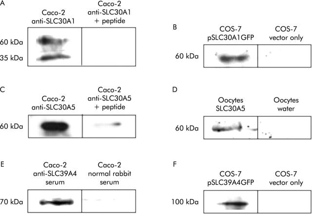
Methods: Subjects took a daily zinc supplement of 25 mg for 14 days in a double blind, placebo controlled, crossover trial. The effect of the supplement on expression in ileal biopsies of the zinc transporters SLC30A1, SLC30A4, SLC30A5, SLC39A1, SLC39A4, and metallothionein was measured by reverse transcription-polymerase chain reaction RT-PCR. Expression of SLC30A1, SLC30A5, and SLC39A4 was also examined by immunoblotting.

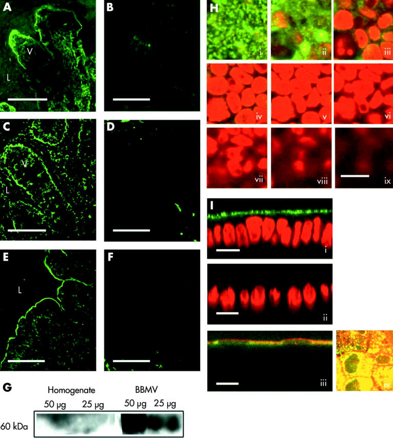
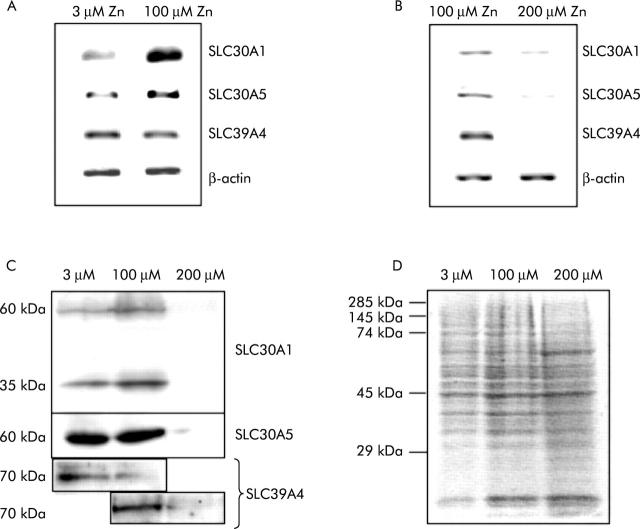
Results: The zinc supplement reduced SLC30A1 mRNA (1.4-fold) together with SLC30A1, SLC30A5, and SLC39A4 protein (1.8-fold, 3.7-fold, and to undetectable levels, respectively) in ileal mucosa and increased metallothionein mRNA (1.7-fold). The supplement had no effect on expression of SLC30A4 or SLC39A1 mRNA. Localisation of SLC30A5 at the apical human enterocyte/colonocyte membrane and also at the apical membrane of Caco-2 cells was demonstrated by immunohistochemistry. Commensurate with these observations in zinc supplemented human subjects, SLC30A1, SLC30A5, and SLC39A4 mRNA and protein were reduced in Caco-2 cells cultured at 200 muM compared with 100 muM zinc.

Conclusions: These observations indicate that, in response to variations in dietary zinc intakes, regulated expression of plasma membrane zinc transporters in the human intestine contributes to maintenance of zinc status.


Figures

Figure 1
Figure 1
Comparison of the expression of zinc related genes in peripheral blood monocytes and biopsies of human small intestine taken either after zinc supplementation or administration of placebo. (A) Reverse transcription-polymerase chain reaction (RT-PCR) products generated using primers specific to the transcripts indicated from poly-A+ RNA prepared from total monocyte RNA pooled from 17 volunteers. (B) RT-PCR products generated using primers specific to the transcripts indicated from poly-A+ RNA prepared from total RNA pooled from single intestinal biopsies from 17 volunteers. For (A) and (B), products generated from poly-A+ RNA prepared from biopsies following administration of placebo or zinc supplement are shown as indicated. PCR was carried out over a non-saturating number of cycles and thus band intensity is representative of the quantity of the specific transcript in the RNA sample. Three independent analyses of the relative expression level of each transcript in the two samples gave comparable results. MT, metallothionein. (C) Analysis by immunoblotting using antipeptide antibodies of expression in pooled biopsies from each of 17 volunteers of the zinc transporters SLC30A1, SLC30A5, and SLC39A4, and of α-tubulin, as indicated. For each sample either 15 μg or 30 μg of protein, as determined by Bradford analysis, were resolved by sodium dodecyl sulphate-polyacrylamide gel electrophoresis prior to blotting. The result of analysis of samples prepared from biopsies following administration of placebo or the zinc supplement are shown as indicated.
Figure 2
Figure 2
Verification of specificity and target protein reactivity of anti-SLC30A1, anti-SLC30A5, and anti-SLC39A4 antipeptide antibodies. (A, B) Immunoblots using anti-SLC30A1 antibody. (C, D) Immunoblots using anti-SLC30A5 antibody. (E, F) Immunoblots using anti-SLC39A4 antibody. Data for Caco-2 cells (A, C, E) were obtained from untransfected cells. Data for COS-7 cells (B, D) were obtained from cells transfected with the plasmid constructs indicated. (A) Protein (7 μg) extracted from Caco-2 cells was run in each lane and the blot was incubated with affinity purified anti-SLC30A1 antibody at a dilution of 1:100 or with the same amount of antibody plus 2.5 μg/ml of the immunising peptide, as indicated. (B) Protein was prepared from COS-7 cells transfected with a plasmid construct (pSLC30A1GFP) from which the C terminal region of SLC30A1 between amino acid residues 334 and 507, inclusive, was expressed as an N terminal fusion to GFP or with vector only (plasmid pEGFPN), as indicated; 20 μg were run in each lane and the blot was incubated with a 1:100 dilution of affinity purified anti-SLC30A1 antibody. (C) Protein (7 μg) extracted from Caco-2 cells was run in each lane and the blot was incubated with affinity purified anti-SLC30A5 antibody at a dilution of 1:100 or with the same amount of antibody plus 2.5 μg/ml of the immunising peptide, as indicated. (D) Protein was prepared from Xenopus laevis oocytes injected with SLC30A5 cRNA or with water (10 of each), as indicated; 20 μg were run in each lane and the blot was incubated with a 1:100 dilution of affinity purified anti-SLC30A5 antibody. (E) Protein (7 μg) extracted from Caco-2 cells was run in each lane and the blot was incubated with a 1:500 dilution of serum from a rabbit treated with the immunising peptide or with a 1:500 dilution of normal rabbit serum, as indicated. (F) Protein was prepared from COS-7 cells transfected with a plasmid construct (pSLC39A4GFP) from which SLC39A4 was expressed as an N terminal fusion to GFP or with vector only (plasmid pEGFPN), as indicated; 20 μg were run in each lane and the blot was incubated with a 1:200 dilution of affinity purified anti-SLC39A4 antibody.
Figure 3
Figure 3
Immunolocalisation of SLC30A5 in human intestine and in Caco-2 cells. Binding of the primary antibody was detected using a FITC conjugated secondary antibody and is shown in green. (A) Section of human jejunum. (C) Section of human ileum. (E) Section of human colon. (B, D, F) Corresponding negative controls stained with secondary antibody only. V, villus; L, lumen; scale bar 50 μm. (G) Result of immunoblotting using anti-SLC30A5 antibody samples of total mucosal homogenate and brush border membrane vesicles (50 μg and 25 μg of protein, as indicated) prepared from human jejunum. A predominant band of approximate 60 kDa molecular weight was observed in the brush border membrane vesicle preparation. (H, I) Staining in Caco-2 cells. Scale bar 25 μm. (H) Consecutive Z sections (parallel to the plane of the monolayer) captured by confocal laser scanning microscopy passing from the apical (i) to the basal (ix) surface of the cell monolayer. SLC30A5 staining is shown in green. Nuclei are stained with propidium iodide and are shown in red. (I) Images (i), (ii), and (iii) are XZ sections (perpendicular to the plane of the monolayer). In images (i) and (ii), nuclei are stained with propidium iodide and are shown in red. Image (iii) shows colocalisation of anti-SLC30A5 immunoreactivity and alkaline phosphatase activity (red). Image (iv) shows colocalisation of anti-SLC30A5 immunoreactivity (green) and alkaline phosphatase activity (red) as a projected Z series viewed from above the cell monolayer.
Figure 4
Figure 4
Regulation by zinc of SLC30A1, SLC30A5, and SLC39A4 expression in Caco-2 cells. (A, B) Reverse transcription-polymerase chain reaction (RT-PCR) products generated from total RNA prepared from Caco-2 cells using primers specific to the transcripts indicated. Products generated from RNA prepared from cells grown for three days at 3, 100, and 200 μM ZnCl2 are shown, as indicated. For each transcript, PCR was carried out over a number of cycles such that the rate of product formation did not reach a plateau, and thus band intensity is representative of the quantity of the specific transcript in the RNA sample. (C) Analysis by immunoblotting using antipeptide antibodies of expression in Caco-2 cells of SLC30A1, SLC30A5, and SLC39A4. Each sample of 7 μg of protein, as determined by Bradford analysis, was resolved by sodium dodecyl sulphate-polyacrylamide gel electrophoresis (SDS-PAGE) prior to blotting. The results of analysis of samples prepared from Caco-2 cells grown for three days at 3, 100, and 200 μM ZnCl2 are shown, as indicated. (D) A replica SDS-PAGE gel, loaded as for the experiments shown in (C), stained with Coomassie brilliant blue to demonstrate equal sample loading. The position of molecular weight standards is indicated.

Similar articles

Cited by

References

    1. Hirst BH. Dietary regulation of intestinal nutrient carriers. Proc Nutr Soc 1993;52:315–24. - PubMed
    1. Ferraris RP, Diamond J. Regulation of intestinal sugar transport. Physiol Rev 1997;77:257–302. - PubMed
    1. Gunshin H, Mackenzie B, Berger UV, et al. Cloning and characterization of a mammalian proton-coupled metal-ion transporter. Nature 1997;388:482–8. - PubMed
    1. Cragg RA, Christie GR, Phillips SR, et al. A novel zinc-regulated human zinc transporter, SLC30A5, is localised to the enterocyte apical membrane. J Biol Chem 2002;277:22789–97. - PubMed
    1. Kambe T, Narita H, Yamaguchi-Iwai Y, et al. Cloning and characterization of a novel mammalian zinc transporter, zinc transporter 5, abundantly expressed in pancreatic beta cells. J Biol Chem 2002;277:19049–55. - PubMed

Publication types

MeSH terms