. 2005 Mar 8;5(1):2.
doi: 10.1186/1472-6874-5-2.

Increased capsaicin receptor TRPV1 in skin nerve fibres and related vanilloid receptors TRPV3 and TRPV4 in keratinocytes in human breast pain

Affiliations

Increased capsaicin receptor TRPV1 in skin nerve fibres and related vanilloid receptors TRPV3 and TRPV4 in keratinocytes in human breast pain

Preethi Gopinath et al. BMC Womens Health. .

Abstract

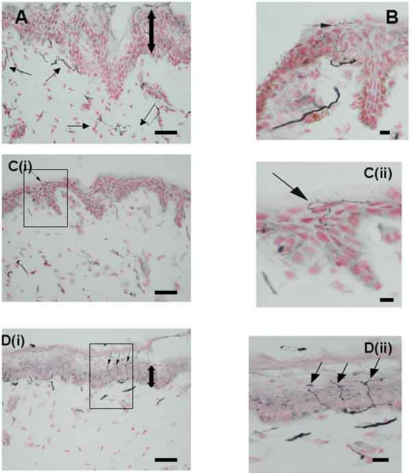
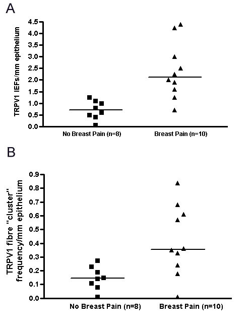
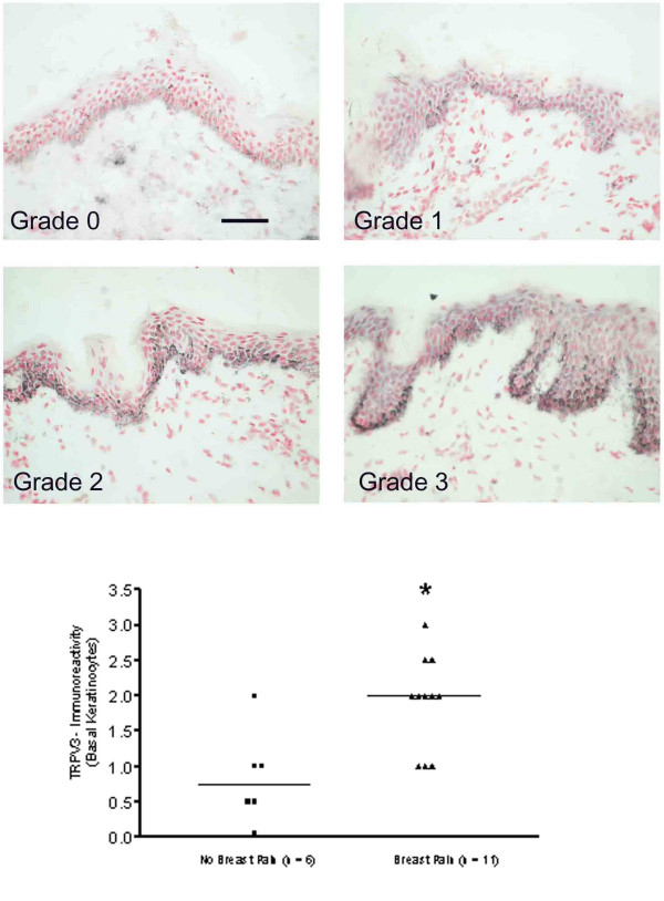
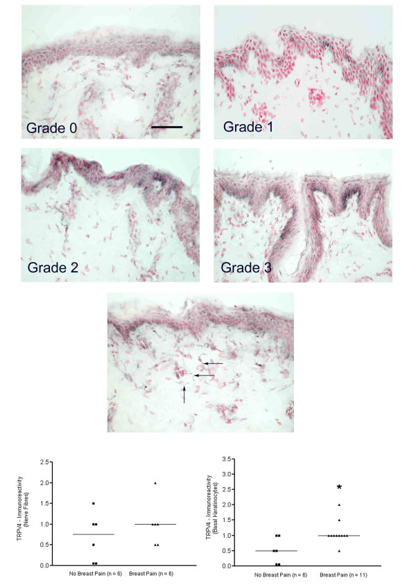
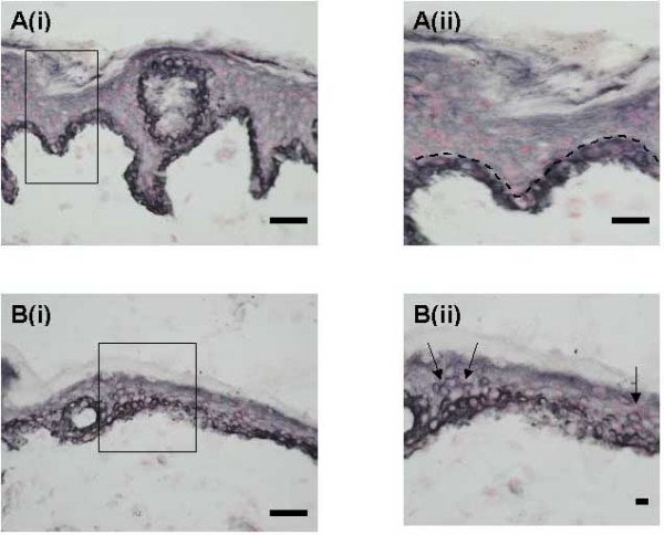
BACKGROUND: Breast pain and tenderness affects 70% of women at some time. These symptoms have been attributed to stretching of the nerves with increase in breast size, but tissue mechanisms are poorly understood. METHODS: Eighteen patients (n = 12 breast reduction and n = 6 breast reconstruction) were recruited and assessed for breast pain by clinical questionnaire. Breast skin biopsies from each patient were examined using immunohistological methods with specific antibodies to the capsaicin receptor TRPV1, related vanilloid thermoreceptors TRPV3 and TRPV4, and nerve growth factor (NGF). RESULTS: TRPV1-positive intra-epidermal nerve fibres were significantly increased in patients with breast pain and tenderness (TRPV1 fibres / mm epidermis, median [range] - no pain group, n = 8, 0.69 [0-1.27]; pain group, n = 10, 2.15 [0.77-4.38]; p = 0.0009). Nerve Growth Factor, which up-regulates TRPV1 and induces nerve sprouting, was present basal keratinocytes: some breast pain specimens also showed NGF staining in supra-basal keratinocytes. TRPV4-immunoreactive fibres were present in sub-epidermis but not significantly changed in painful breast tissue. Both TRPV3 and TRPV4 were significantly increased in keratinocytes in breast pain tissues; TRPV3, median [range] - no pain group, n = 6, 0.75 [0-2]; pain group, n = 11, 2 123, p = 0.008; TRPV4, median [range] - no pain group, n = 6, [0-1]; pain group, n = 11, 1 [0.5-2], p = 0.014). CONCLUSION: Increased TRPV1 intra-epidermal nerve fibres could represent collateral sprouts, or re-innervation following nerve stretch and damage by polymodal nociceptors. Selective TRPV1-blockers may provide new therapy in breast pain. The role of TRPV3 and TRPV4 changes in keratinocytes deserve further study.


Figures

Figure 1
Figure 1
TRPV1-immunoreactive nerve fibres in breast skin. (A) Normal, control skin : TRPV1-immunoreactive nerve fibres (small arrows) in the sub-epidermis. (B) Painful skin (macromastia patient): intra-epidermal, TRPV1-immunoreactive nerve fibre (arrow) deriving from a large, sub-epidermal fascicle and extending to the stratum corneum. (Ci) Painful skin (breast reconstruction patient): TRPV1-immunoreactive intra-epidermal fibres passing along the junction between the epidermis and stratum corneum (Cii-enlarged area from Ci). (Di) Painful skin (macromastia patient): multiple branching, TRPV1-immunoreactive intra-epidermal nerve fibres (arrows) extending to the stratum corneum (Dii-enlarged area from Di). Large double arrows indicate relative epidermal thickness. Scale bars: A, B, C(i), D(i) = 50 μm; C(ii), D(ii) = 10 μm.
Figure 2
Figure 2
Quantification of TRPV1-immunoreactive, intra-epidermal fibres and breast pain. Scattergrams show TRPV1-immunoreactivity in (A) intra-epidermal fibres and (B) intra-epidermal fibre "clusters" in patients with and without breast pain.
Figure 3
Figure 3
TRPV3-immunoreactivity in breast skin. TOP PANELS: TRPV3-immunoreactive keratinocytes mostly in basal layer and graded from grade 0/negative (top left) to grade 3/strong staining (bottom right). Scale bar = 100 μm BOTTOM PANEL: Scattergram showing grading assessment and a significant increase (*P < 0.05) of TRPV3 immunoreactivity in patients with pain.
Figure 4
Figure 4
TRPV4-immunoreactivity in breast skin. TOP FOUR PANELS: TRPV4-immunoreactivity in keratinocytes mostly at the cell membrane and graded from grade 0/negative (top left) to grade 3/strong staining (bottom right). Scale bar = 100 μm MIDDLE PANEL: Fine, sub-epidermal, TRPV4-immunoreactive fibres (arrows). Scale bar = 100 μm BOTTOM PANELS: Scattergrams showing grading assessment of sub-epidermal fibres (left panel) and keratinocytes (right panel) significantly increased (*P < 0.05) in patients with pain.
Figure 5
Figure 5
NGF-immunoreactivity in breast skin. (Ai) Normal, control skin: NGF-immunoreactive basal keratinocytes. (Aii)-enlarged area from Ai showing NGF confined to single layer of keratinocytes. (Bi) Painful skin (macromastia patient): NGF-immunoreactive basal and supra-basal (Bii-arrows) keratinocytes in skin with thin epidermis.

Similar articles

Cited by

References

    1. Wallace MS, Wallace AM, Lee J, Dobke MK. Pain after breast surgery: a survey of 282 women. Pain. 1996;66:195–205. doi: 10.1016/0304-3959(96)03064-3. - DOI - PubMed
    1. Riley JL, Robinson ME, Wise EA, Price DD. A meta-analytic review of pain perception across the menstrual cycle. Pain. 1999;81:225–235. doi: 10.1016/S0304-3959(98)00258-9. - DOI - PubMed
    1. Holdcroft A. Females and their variability. Anaesthesia. 1997;52:931–934. doi: 10.1111/j.1365-2044.1997.265-az0393.x. - DOI - PubMed
    1. Morin C, Bushnell MC. Temporal and qualitative properties of cold pain and heat pain: a psychophysical study. Pain. 1998;74:67–73. doi: 10.1016/S0304-3959(97)00152-8. - DOI - PubMed
    1. Robinson JE, Short RV. Changes in breast sensitivity at puberty, during the menstrual cycle, and at parturition. Br Med J. 1977;1:1188–1191. - PMC - PubMed