Comparative Study
. 2005 May 6;280(18):18536-42.
doi: 10.1074/jbc.M413224200. Epub 2005 Mar 8.

Manganese superoxide dismutase protects against 6-hydroxydopamine injury in mouse brains

Affiliations
Comparative Study

Manganese superoxide dismutase protects against 6-hydroxydopamine injury in mouse brains

Jason Callio et al. J Biol Chem. .

Abstract

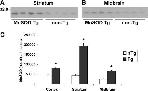
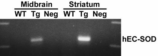
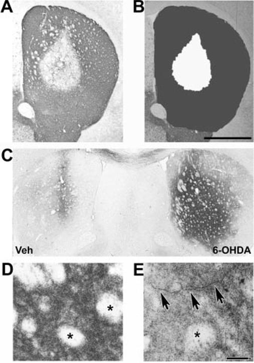
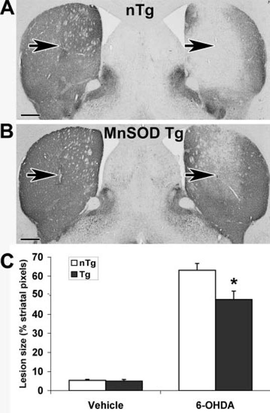
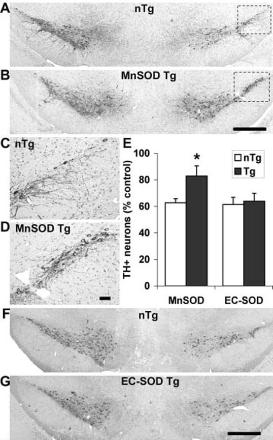
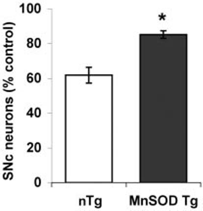
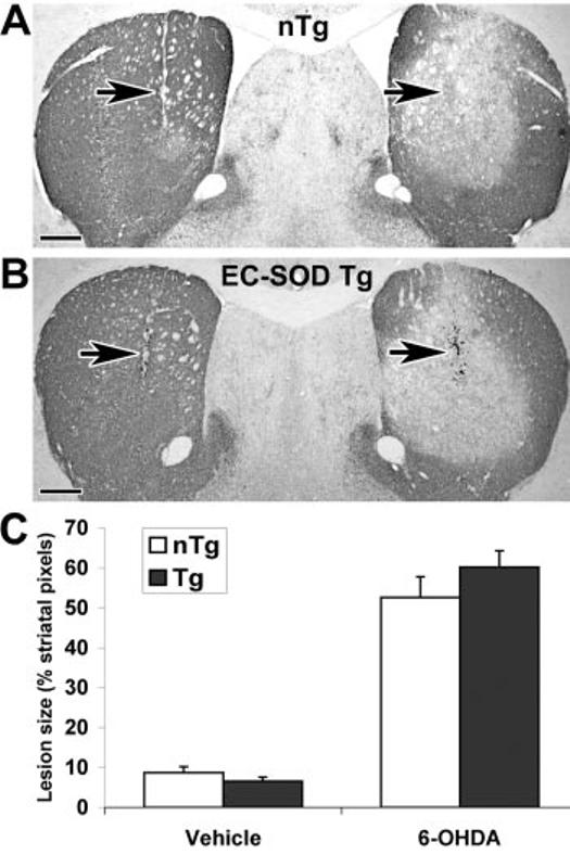
Dopaminergic neurons of the substantia nigra are susceptible to toxin-based insults. Intrastriatal injection of 6-hydroxydopamine results in selective toxicity to these neurons. A mechanistic role for reactive oxygen species is supported by observations that antioxidants confer protection from 6-hydroxydopamine. Although cell culture studies have suggested extracellular or nonmitochondrial mechanisms in 6-hydroxydopamine toxicity, the compartmentalization of oxidative injury mechanisms is incompletely defined in vivo. Transgenic mice overexpressing mitochondrial manganese superoxide dismutase or extracellular superoxide dismutase received unilateral intrastriatal injections of 6-hydroxydopamine. Mice that overexpress manganese superoxide dismutase showed significantly smaller striatal lesions than littermate controls. There were no differences in nonspecific striatal injury associated with contralateral vehicle injection. Manganese superoxide dismutase overexpression also protected against loss of neuronal cell bodies in the substantia nigra. In contrast, mice overexpressing extracellular superoxide dismutase showed no protection from 6-hydroxydopamine toxicity in either brain region. Protection of the nigrostriatal system by overexpression of manganese superoxide dismutase supports a role for mitochondrially derived superoxide in 6-hydroxydopamine toxicity. Mitochondrial oxidative stress appears to be a common mechanism among diverse models of Parkinson disease, whether involving toxins, mutated genes, or cybrid cells containing patient mitochondria. Antioxidant therapies that target this subcellular compartment may prove promising.


Figures

Fig.1
Fig.1
Western blot analysis shows increased Mn-SOD expression in the nigrostriatal system. Each lane represents dissected (A) striata or (B) ventral midbrains from three or four mice of the same genotype which were pooled during homogenization (15 μg of total protein loaded). Note the increased Mn-SOD protein expression in the Tg mice in both brain regions. C, densitometry of Mn-SOD bands from the cortex, striatum, and midbrains of Mn-SOD Tg (dark bars) and non-Tg (nTg) littermates (white bars) confirms significant protein over-expression in all brain regions. Values represent the mean ± S.E. * p < 0.05 compared with non-Tg controls by Student's t test for paired samples.
Fig. 2
Fig. 2
RT-PCR studies show expression of the human EC-SOD transgene in the nigrostriatal system. RT-PCR analysis for human EC-SOD expression in the ventral midbrain and striatum of wild-type (WT) and EC-SOD Tg mice. Note the expression of the human trans-gene in Tg mice. The band is not amplified in control reactions using RNase-pretreated Tg RNA (Neg).
Fig. 3
Fig. 3
Analysis of striatal lesions elicited by intrastriatal injection of 6-OHDA. Wild-type B6C3 mice received unilateral injections of 6-OHDA free base in their right striata. After 7 days, the animals were sacrificed and their striata processed for TH immunohistochemistry. The 0.5–2.5-μg doses resulted in subsaturating lesions amenable to volumetric analysis, whereas 5-μg doses produced saturating lesions. A, representative section reveals loss of TH immunoreactivity extending from the center of the injection site. B, after digital image capture, the striata were defined as regions of interest (dark gray), and the size of the lesion (white) was determined using the autothresholding function of MetaMorph software. For each mouse, this was performed for the vehicle-injected and the 6-OHDA-injected sides on five uniformly spaced levels spanning the rostral-caudal extent of the striatum. Scale bar, 1,000 μm. C, amino cupric silver degeneration stain of a representative mouse reveals intense staining of the 6-OHDA lesioned right striatum at 1 day after injection. In contrast, cupric silver staining was limited to the region of the needle track on the vehicle-injected left side. D, intense punctate staining of striatal termini in a region distant to the needle track at 1 day after 6-OHDA injection. Note sparing of the crossing white matter bundles that do not contain dopaminergic processes (asterisks). E, axonal degeneration is evident at 4 days after 6-OHDA injection (arrows). Scale bar, 40 μm.
Fig. 4
Fig. 4
Tg mice overexpressing Mn-SOD are significantly protected from 6-OHDA injury to the striatum (n = 9 mice/group). Mn-SOD Tg mice and non-Tg littermates received intrastriatal injections of 2.5 μg of 6-OHDA free base on the right and an equivalent volume of vehicle on the left. Representative TH-immunostained brain sections at the level of the needle track (arrows) are shown for a non-Tg (nTg) littermate (A) and Mn-SOD Tg mouse (B). Five sections representing the rostral-caudal extent of the striatum were analyzed as described under “Experimental Procedures” and in Fig. 3B. Summated lesion pixels were expressed as a percent of summated total striatal pixels from the five levels. C, the average lesion sizes ± S.E. are shown for the vehicle-injected left side and the 6-OHDA-injected right side of non-Tg littermate controls (white bars) and Mn-SOD Tg mice (dark bars). * p < 0.05 by analysis of variance followed by Student's t test with Bonferroni correction. Scale bar, 500 μm.
Fig.5
Fig.5
Overexpression of Mn-SOD, but not EC-SOD, confers protection from 6-OHDA-induced TH+ neuron loss in the substantia nigra. Tg mice overexpressing Mn-SOD (n = 9 mice/group) or those overexpressing EC-SOD (n = 7 mice/group) were injected with 6-OHDA or vehicle as described under “Experimental Procedures.” Midbrain sections were stained for TH/hematoxylin and analyzed as described under “Experimental Procedures.” There were no significant differences between Tg and littermate controls in the number of TH+ SNc neuronal profiles on the vehicle-injected left sides (see “Results”). Representative sections for Mn-SOD Tg mice (B) show significant preservation of TH+ SNc neurons on the 6-OHDA-injected right side compared with non-Tg (nTg) littermate controls (A). The lateral SNc of the lesioned side for non-Tg (C) and Mn-SOD Tg (D) mice are shown at higher magnification. In contrast, no differences were observed in TH+ SNc neuron numbers between EC-SOD Tg mice (G) and their control littermates (F). The data obtained from non-Tg (white bars) and Tg (dark bars) mice of both genotypes, expressed as a percent of the vehicle-injected control side, are graphed for comparison (E). * p < 0.05 by analysis of variance followed by Student's t test with Bonferroni correction. Scale bars, 500 μm for A, B, F and G and 50 μm for C and D.
Fig.6
Fig.6
Overexpression of Mn-SOD confers protection from 6-OHDA-induced SNc neuron cell loss in the substantia nigra. The total number of SNc neurons in midbrain sections from Mn-SOD Tg mice and their littermate controls (nTg) was analyzed by counting hematoxylin-stained neuronal nuclei. The data obtained from non-Tg (white bar) and Mn-SOD Tg (dark bar) mice, expressed as a percent of the vehicle-injected control side, are graphed for comparison. * p < 0.05 by Student's t test.
Fig.7
Fig.7
Tg mice overexpressing EC-SOD are not protected from 6-OHDA injury to the striatum (n = 7 mice/group). EC-SOD Tg mice and non-Tg littermates received intrastriatal injections of 2.5 μg of 6-OHDA free base on the right and an equivalent volume of vehicle on the left. Representative TH-immunostained brain sections at the level of the needle track (arrows) are shown for a non-Tg (nTg) littermate (A) and EC-SOD Tg mouse (B). Five sections representing the rostralcaudal extent of the striatum were analyzed as described under “Experimental Procedures.” Scale bars, 500 μm. C, the average lesion size ± S.E. are shown for the vehicle-injected left side and the 6-OHDA-injected right side of non-Tg littermate controls (white bars) and ECSOD Tg mice (dark bars).

Similar articles

Cited by

References

    1. Oury TD, Ho Y-S, Piantadosi CA, Crapo JD. Proc. Natl. Acad. Sci. U. S. A. 1992;89:9715–9719. - PMC - PubMed
    1. Fullerton HJ, Ditelberg JS, Chen SF, Sarco DP, Chan PH, Epstein CJ, Ferriero DM. Ann. Neurol. 1998;44:357–364. - PubMed
    1. Klivenyi P, Clair D, Wermer M, Yen HC, Oberley T, Yang L, Flint Beal M. Neurobiol. Dis. 1998;5:253–258. - PubMed
    1. Oury TD, Day BJ, Crapo JD. Lab. Invest. 1996;75:617–636. - PubMed
    1. Oury TD, Piantadosi CA, Crapo JD. J. Biol. Chem. 1993;268:15394–15398. - PubMed

Publication types

LinkOut - more resources