

Conformational flexibility revealed by the crystal structure of a crenarchaeal RadA
- PMID: 15755748
- PMCID: PMC1062875
- DOI: 10.1093/nar/gki288
Conformational flexibility revealed by the crystal structure of a crenarchaeal RadA
Abstract
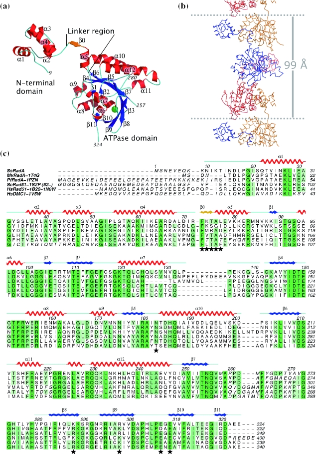
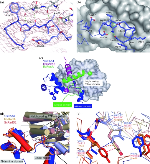
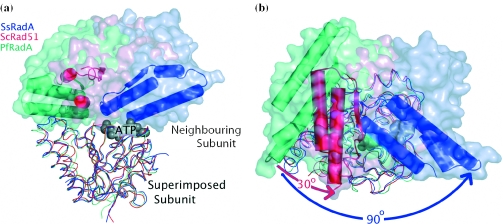
Homologous recombinational repair is an essential mechanism for repair of double-strand breaks in DNA. Recombinases of the RecA-fold family play a crucial role in this process, forming filaments that utilize ATP to mediate their interactions with single- and double-stranded DNA. The recombinase molecules present in the archaea (RadA) and eukaryota (Rad51) are more closely related to each other than to their bacterial counterpart (RecA) and, as a result, RadA makes a suitable model for the eukaryotic system. The crystal structure of Sulfolobus solfataricus RadA has been solved to a resolution of 3.2 A in the absence of nucleotide analogues or DNA, revealing a narrow filamentous assembly with three molecules per helical turn. As observed in other RecA-family recombinases, each RadA molecule in the filament is linked to its neighbour via interactions of a short beta-strand with the neighbouring ATPase domain. However, despite apparent flexibility between domains, comparison with other structures indicates conservation of a number of key interactions that introduce rigidity to the system, allowing allosteric control of the filament by interaction with ATP. Additional analysis reveals that the interaction specificity of the five human Rad51 paralogues can be predicted using a simple model based on the RadA structure.
Figures
Similar articles
-
Crystal structure of the left-handed archaeal RadA helical filament: identification of a functional motif for controlling quaternary structures and enzymatic functions of RecA family proteins.Nucleic Acids Res. 2007;35(6):1787-801. doi: 10.1093/nar/gkl1131. Epub 2007 Feb 28. Nucleic Acids Res. 2007. PMID: 17329376 Free PMC article.
-
Three new structures of left-handed RADA helical filaments: structural flexibility of N-terminal domain is critical for recombinase activity.PLoS One. 2009;4(3):e4890. doi: 10.1371/journal.pone.0004890. Epub 2009 Mar 19. PLoS One. 2009. PMID: 19295907 Free PMC article.
-
Structural and functional characterisation of a conserved archaeal RadA paralog with antirecombinase activity.J Mol Biol. 2009 Jun 19;389(4):661-73. doi: 10.1016/j.jmb.2009.04.060. Epub 2009 May 3. J Mol Biol. 2009. PMID: 19414020 Free PMC article.
-
Right or left turn? RecA family protein filaments promote homologous recombination through clockwise axial rotation.Bioessays. 2008 Jan;30(1):48-56. doi: 10.1002/bies.20694. Bioessays. 2008. PMID: 18081011 Review.
-
What is the structure of the RecA-DNA filament?Curr Protein Pept Sci. 2004 Apr;5(2):73-9. doi: 10.2174/1389203043486883. Curr Protein Pept Sci. 2004. PMID: 15078218 Review.
Cited by
-
Divalent metal cofactors differentially modulate RadA-mediated strand invasion and exchange in Saccharolobus solfataricus.Biosci Rep. 2023 Feb 27;43(2):BSR20221807. doi: 10.1042/BSR20221807. Biosci Rep. 2023. PMID: 36601994 Free PMC article.
-
Crystal structure of the left-handed archaeal RadA helical filament: identification of a functional motif for controlling quaternary structures and enzymatic functions of RecA family proteins.Nucleic Acids Res. 2007;35(6):1787-801. doi: 10.1093/nar/gkl1131. Epub 2007 Feb 28. Nucleic Acids Res. 2007. PMID: 17329376 Free PMC article.
-
Structural and functional analyses of five conserved positively charged residues in the L1 and N-terminal DNA binding motifs of archaeal RADA protein.PLoS One. 2007 Sep 12;2(9):e858. doi: 10.1371/journal.pone.0000858. PLoS One. 2007. PMID: 17848989 Free PMC article.
-
TWINKLE and Other Human Mitochondrial DNA Helicases: Structure, Function and Disease.Genes (Basel). 2020 Apr 9;11(4):408. doi: 10.3390/genes11040408. Genes (Basel). 2020. PMID: 32283748 Free PMC article. Review.
-
Three new structures of left-handed RADA helical filaments: structural flexibility of N-terminal domain is critical for recombinase activity.PLoS One. 2009;4(3):e4890. doi: 10.1371/journal.pone.0004890. Epub 2009 Mar 19. PLoS One. 2009. PMID: 19295907 Free PMC article.
References
-
- Bianco P.R., Tracy R.B., Kowalczykowski S.C. DNA strand exchange proteins: a biochemical and physical comparison. Front. Biosci. 1998;3:D570–D603. - PubMed
-
- Lusetti S.L., Cox M.M. The bacterial RecA protein and the recombinational DNA repair of stalled replication forks. Annu. Rev. Biochem. 2002;71:71–100. - PubMed
-
- White M.F. Archaeal DNA repair: paradigms and puzzles. Biochem. Soc. Trans. 2003;31:690–693. - PubMed
-
- Komori K., Miyata T., Daiyasu H., Toh H., Shinagawa H., Ishino Y. Domain analysis of an archaeal RadA protein for the strand exchange activity. J. Biol. Chem. 2000;275:33791–33797. - PubMed
Publication types
MeSH terms
Substances
LinkOut - more resources
Full Text Sources
Other Literature Sources
Research Materials
