. 2005 Mar 8;33(5):1487-502.
doi: 10.1093/nar/gki290. Print 2005.

Characterizing the function and structural organization of the 5' tRNA-like motif within the hepatitis C virus quasispecies

Affiliations

Characterizing the function and structural organization of the 5' tRNA-like motif within the hepatitis C virus quasispecies

Maria Piron et al. Nucleic Acids Res. .

Abstract

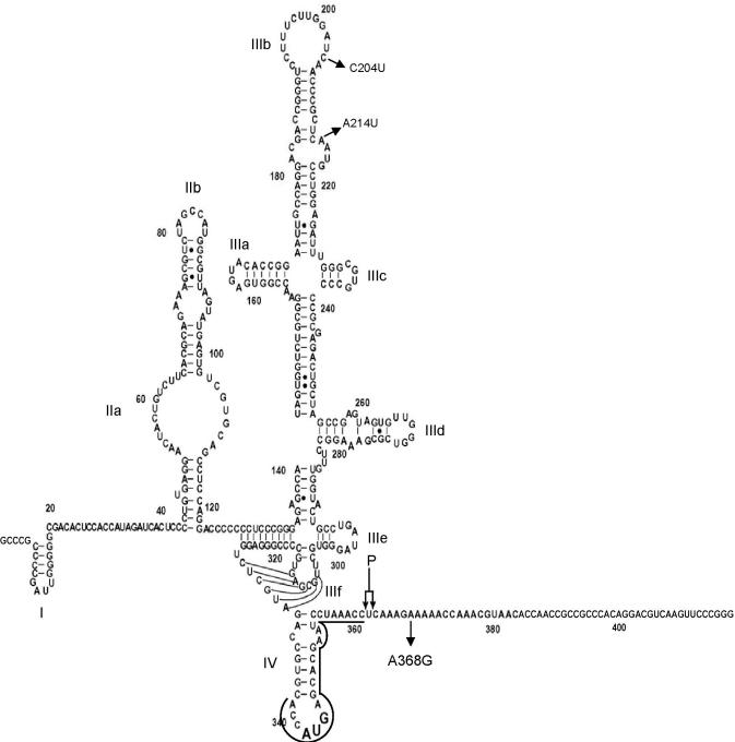
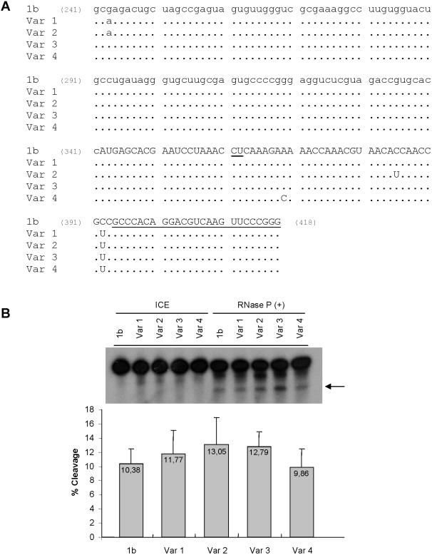
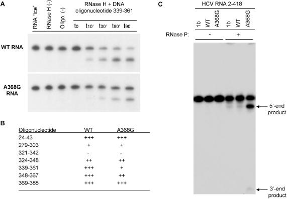
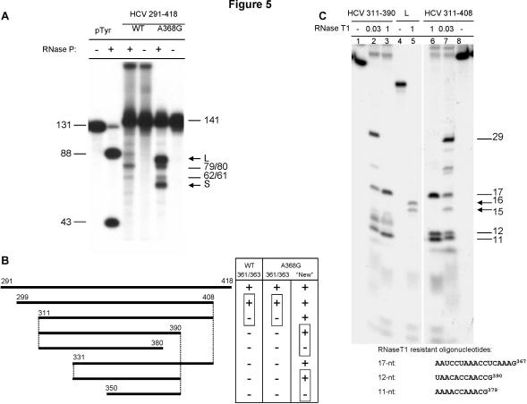
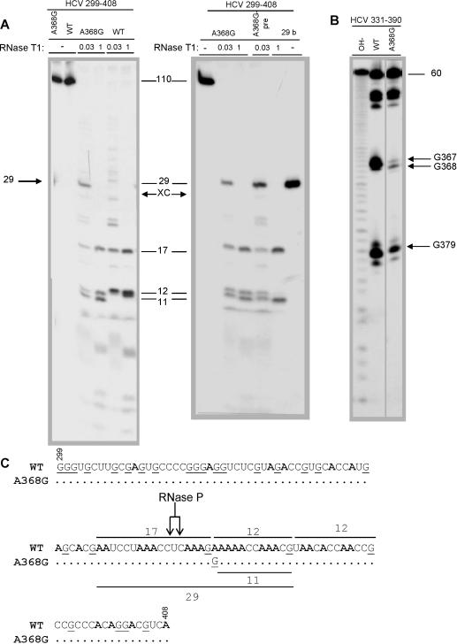
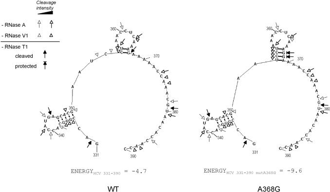
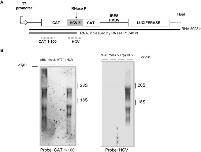
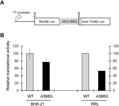
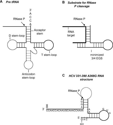
Hepatitis C virus (HCV) RNA is recognized and cleaved in vitro by RNase P enzyme near the AUG start codon. Because RNase P identifies transfer RNA (tRNA) precursors, it has been proposed that HCV RNA adopts structural similarities to tRNA. Here, we present experimental evidence of RNase P sensitivity conservation in natural RNA variant sequences, including a mutant sequence (A368-G) selected in vitro because it presented changes in the RNA structure of the relevant motif. The variation did not abrogate the original RNase P cleavage, but instead, it allowed a second cleavage at least 10 times more efficient, 4 nt downstream from the original one. The minimal RNA fragment that confers sensitivity to human RNase P enzyme was located between positions 299 and 408 (110 nt). Therefore, most of the tRNA-like domain resides within the viral internal ribosome entry site (IRES) element. In the variant, in which the mutation stabilizes a 4 nt stem-loop, the second cleavage required a shorter (60 nt) substrate, internal to the minimal fragment substrate, conforming a second tRNA-like structure with similarities to a 'Russian-doll' toy. This new structure did not impair IRES activity, albeit slightly reduced the efficiency of translation both in vitro and in transfected cells. Conservation of the original tRNA-like conformation together with preservation of IRES activity points to an essential role for this motif. This conservation is compatible with the presence of RNA structures with different complexity around the AUG start codon within a single viral population (quasispecies).


Figures

Figure 1
Figure 1
Diagram of genotype 1b HCV IRES RNA secondary structure (sequence 2–418 used in this work) modified from (25). Substitutions C204U and A214U in the WT sequence are indicated; the A368G sequence only differs from the WT sequence by the A368G mutation. A double arrow indicates RNase P cleavage site (361–363). A line between nucleotides 339 and 361 depicts the hybridization site of a DNA primer used for in vitro selection in Figure 2. Structural sub-domains are indicated from I to IV.
Figure 2
Figure 2
Schematic representation of in vitro selection procedure for isolating natural HCV IRES RNA structure variants using RNase H/DNA oligonucleotide-mediated cleavage of HCV mRNA. See Materials and Methods for details.
Figure 3
Figure 3
RNase P sensitivity of HCV RNA natural variants bearing substitutions in the RNase P cleavage region. (A) Sequence alignment of nucleotides 241–418 of cloned HCV variants. 1b represents the genotype 1b reference sequence and variants 1–4, four HCV variants isolated through cloning and sequencing of 2–418 region of HCV RNA (lower case, HCV 5′ NCR, upper case, HCV core coding region). RNase P cleavage site is underlined at positions 361–363. The position of the 3′ primer used for PCR and cloning is underlined (394–418). (B) Autoradiogram of RNase P in vitro cleavage assay of labeled 2–418 HCV RNAs. Variants 1–4 were incubated with RNase P, in parallel to 1b HCV RNA control, 1 h 30 min at 30°C. ICE indicates the input RNA, not incubated. Reaction products were fractionated in denaturing polyacrylamide–UREA gel electrophoresis followed of X-ray film exposure. The arrow indicates the larger cleavage product. Experiment was repeated as a triplicate and quantified with a Radioisotopic Image Analyser FLA-5000 (Fuji). The table below the autoradiogram indicates the average cleavage of each RNA determined as the ratio: products/products + uncleaved RNA.
Figure 4
Figure 4
Enhanced accessibility to RNase P of the in vitro selected HCV RNA variant partially resistant to RNase H. (A) Time course DNA-mediated RNase H cleavage of the [α-32P]GTP-labeled WT RNA compared to the A368G variant RNA (nucleotides 2–418), selected as indicated in Figure 2 using a DNA oligonucleotide complementary to nucleotides 339–361 of HCV RNA. Products were analyzed by denaturing PAGE (4%) and visualized by autoradiography. The kinetic study (0–90 min) shows the resistant phenotype associated with the mutant natural variant (estimated at a 3-fold effect). (B) Relative sensitivities of WT and A368G 2–418 HCV RNA to DNA-mediated RNase H cleavage. The oligonucleotide column indicates the positions where DNA oligonucleotides hybridize to RNA. +++: 75–100% of cleavage after 90 min, ++: 50–75%. +: 25–50%, −: 0–25%. (C) In vitro human RNase P cleavage of HCV 2–418 RNA. Lane 1b is the control genotype 1b HCV clone previously used in the laboratory to study RNase P cleavage (10) and represents here the cleavage efficiency and the product size control, lane WT is the HCV predominant sequence of the patient studied here, and lane A368G is the selected mutant sequence. RNase P (+) and (−) are, respectively, reactions with and without RNase P. Products were analyzed by denaturing PAGE (4%) and visualized by autoradiography. The position of the two 5′ and 3′ end RNase P cleavage products is indicated on the right.
Figure 5
Figure 5
Identification of the minimal substrate for RNase P cleavage in the in vitro selected HCV RNA variant. (A) Comparison of the RNase P cleavage pattern of HCV WT and A368G. 32P-labeled natural substrate for human RNase P (pTyr), HCV WT and A368G 291–418 RNAs were subjected (+) or not (−) to in vitro RNase P cleavage and analyzed on a 10% denaturing acrylamide gel. The arrows indicate the new cleavage products observed in A368G RNA, L (Large) and S (Short). Numbers indicate the length in nucleotides of RNA fragments. (B) Delimitation of the human RNase P minimal substrates in HCV WT or A368G RNA IRES. DNA templates were obtained by PCR, in vitro transcribed, and subjected to in vitro RNase P cleavage. Positions of RNase P cleavage are indicated as 361/363 for the previously described cleavage site, and as ‘new’ for the cleavage site described in this work. (C) Identification of the RNase P cleavage products. HCV mutant RNA 311–390 was treated with RNase P. Then, the 5′ end large cleavage product (L) was gel purified and subjected (lane 5) or not (lane 4) to total cleavage by RNase T1. Reaction products were analyzed on a 20% denaturing gel and compared to the HCV mutant RNA 311–390, also subjected to RNase T1 digestion (lanes 1, 2 and 3). RNase T1 cleavage fragments were run in parallel to those obtained at the same time from the 311–408 control RNA that allow the identification of the 11, 12 and 17 nt bands (lanes 6, 7 and 8). Numbers on the right indicate the length in nucleotides of RNA fragments. The table below the gel depicts the sequence of the 11, 12 and 17 nt HCV mutant RNA RNase T1 resistant oligonucleotides.
Figure 6
Figure 6
Enzymatic structural probing of the minimal fragment recognized by RNase P. (A) Comparison of RNase T1 cleavage pattern of WT and A368G uniformly ATP-labeled HCV RNAs (299–408). Left panel: A368G and WT RNA without RNase T1, or incubated with 0.03 or 1 μg/μl of RNase T1 during 20 min at 37°C in RNase P incubation buffer (10 mM HEPES–KOH, pH 7.5, 10 mM MgOAc, 100 mM NH4OAc). Right panel: ‘A368G’: control reaction of mutant RNA incubated with 0.03 μg/μl or 1 μg/μl of RNase T1. ‘A368G-pre’: aliquot of HCV mutant 299–408 RNA cleaved by 0.03 μg/μl RNase T1, before gel purification of the 29 nt product. ‘29 b’: gel-purified 29 nt product treated with or without 1 μg/μl RNase T1. Products were analyzed in denaturing PAGE (20%) and visualized by autoradiography. XC indicates xylene cyanol dye position (separates as a 28 nt fragment in 20% acrylamide gel); numbers in column indicate the length in nucleotides of RNA fragments. (B) RNase T1 partial digestion of 60 nt HCV 331–390 WT and A368G mutant RNAs. 3′ end-labeled RNAs were incubated with 0.001 μg/μl RNase T1, 20 min at 37°C and electrophoresed in a 20% acrylamide gel in parallel with one of the transcripts subjected to partial alkaline hydrolysis (OH-). (C) Diagram of RNase T1 cleavage products in WT and A368G HCV RNA sequences (299–408). The 32P-labeled A residues appear in bold, G residues recognized by RNase T1 are underlined, and position of the described RNase P cleavage site in genotype 1b RNA is indicated. Numbers and lines indicate the largest predicted fragments. Position of the 29 nt product is also underlined.
Figure 7
Figure 7
Summary of secondary structure probing of HCV 331–390 (WT and A368G) RNAs. Sensitivity to various RNases is indicated on the predicted RNA structure. HCV331–390 WT structure was forced for base pairing between CU355–356 and AG366–367. HCV331–390 A368G was predicted by RNA structure 3.5 program (34) (only one structure was predicted). Arrows indicate cleavages by single-strand-specific RNases T1 and A, whereas triangles show cleavage by double-strand-specific RNase V1. Nucleotide numbering is used as in Figure 1 (5′–3′ orientation).
Figure 8
Figure 8
RNase P does not cleave HCV 5′ tRNA-like motif in the cytoplasm of transfected cells. (A) Schematic representation of the plasmid used to test the RNase P cleavage in the cytoplasm. HCV minimal domain (WT) cleaved in vitro by RNase P (HCV 299–408) was inserted in the coding sequence of CAT in the pBIC plasmid. The expected size of the RNA fragments, if cleaved by RNase P, is indicated. Positions of hybridization of CAT 1–100 and HCV299–408 probes are indicated in dotted line (CAT 1–100 and HCV, respectively). (B) Northern blot analysis after transfection of BHK-21 cells with pBIC-HCV. BHK-21 cells were transfected with pBIC plasmids containing (HCV) or not (pBIC) the HCV target for RNase P in vitro cleavage. pBIC plasmids were linearized by HpaI before transfection. Controls were cells not transfected/infected (mock) or cells not infected [VT7(−)]. Total RNA was extracted from the cells at 16 h post-transfection, electrophoresed, transfered to a nylon membrane and hybridized with a pBIC-specific probe (CAT) or HCV-specific probe (HCV). The position of the ribosomal 28S and 18S RNA markers is indicated on the right.
Figure 9
Figure 9
Effect of A368G mutation on IRES activity. (A) Diagram of the bicistronic plasmids used for the translation assays. The HCV RNA sequence is shown in a gray box (2–418). The first 26 amino acids of HCV core protein are fused in frame with the firefly luciferase. (B) Comparison of WT and A368G HCV IRES translation efficiency. IRES activity was assessed by measuring the ratio of Firefly Luciferase to Renilla Luciferase produced from plasmids transfected in BHK-21 cells or from in vitro transcribed RNA in rabbit reticulocyte lysate (RRL).
Figure 10
Figure 10
Comparison of HCV A368G RNA structure to that of human RNase P minimal substrate. (A) Diagram of pre-tRNA cloverleaf structure. An arrow indicates human RNase P cleavage site. Plain lines indicate the minimal structure necessary for RNase P recognition: acceptor stem, T stem–loop and D stem. (B) Diagram of a minimized external guide sequence (3/4 EGS) hybridized to a target RNA and recognized by human RNase P. (C) Representation of HCV A368G RNA structure (nucleotides 331–390, from Figure 8) to allow a comparison with a 3/4 EGS. The arrow indicates RNase P cleavage site in HCV A368G RNA.

Similar articles

Cited by

References

    1. Martell M., Esteban J.I., Quer J., Genesca J., Weiner A., Esteban R., Guardia J., Gomez J. Hepatitis C virus (HCV) circulates as a population of different but closely related genomes: quasispecies nature of HCV genome distribution. J. Virol. 1992;66:3225–3229. - PMC - PubMed
    1. Domingo E., Holland J.J. Mutation rates and rapid evolution of RNA viruses. In: Morse S.S., editor. Evolutionary Biology of Viruses. New York: Raven Press; 1994. pp. 161–184.
    1. Schuster P. Genotypes with phenotypes: adventures in an RNA toy world. Biophys. Chem. 1997;66:75–110. - PubMed
    1. Tsukiyama-Kohara K., Iizuka N., Kohara M., Nomoto A. Internal ribosome entry site within hepatitis C virus RNA. J. Virol. 1992;66:1476–1483. - PMC - PubMed
    1. Honda M., Ping L.H., Rijnbrand R.C., Amphlett E., Clarke B., Rowlands D., Lemon S.M. Structural requirements for initiation of translation by internal ribosome entry within genome-length hepatitis C virus RNA. Virology. 1996;222:31–42. - PubMed

Publication types

Associated data