

Effects of titanium particle size on osteoblast functions in vitro and in vivo
- PMID: 15755807
- PMCID: PMC555523
- DOI: 10.1073/pnas.0500693102
Effects of titanium particle size on osteoblast functions in vitro and in vivo
Abstract
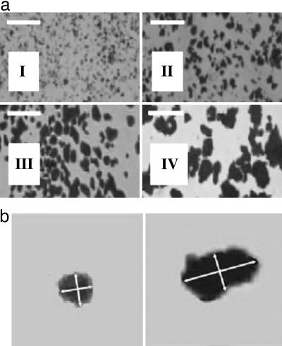
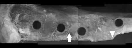
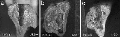
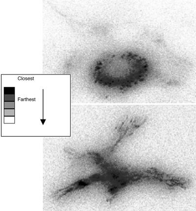
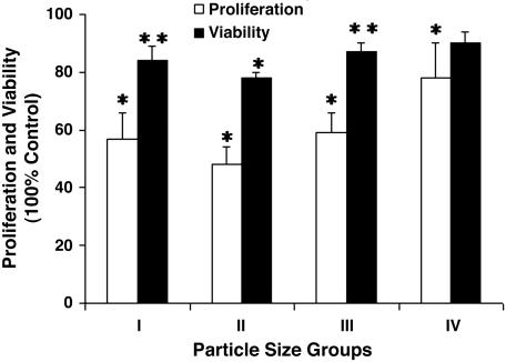
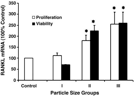
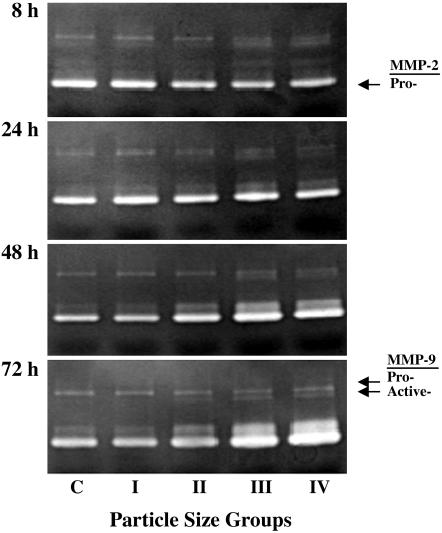
The formation of titanium (Ti)-wear particles during the lifetime of an implant is believed to be a major component of loosening due to debris-induced changes in bone cell function. Radiographic evidence indicates a loss of fixation at the implant-bone interface, and we believe that the accumulation of Ti particles may act on the bone-remodeling process and impact both long- and short-term implant-fixation strengths. To determine the effects of various sizes of the Ti particles on osteoblast function in vivo, we measured the loss of integration strength around Ti-pin implants inserted into a rat tibia in conjunction with Ti particles from one of four size-groups. Implant integration is mediated primarily by osteoblast adhesion/focal contact pattern, viability, proliferation and differentiation, and osteoclast recruitment at the implant site in vivo. This study demonstrates the significant attenuation of osteoblast function concurrent with increased expression of receptor activator of nuclear factor kappaB ligand (RANKL), a dominant signal for osteoclast recruitment, which is regulated differentially, depending on the size of the Ti particle. Zymography studies have also demonstrated increased activities of matrix metalloproteinases (MMP) 2 and 9 in cells exposed to larger Ti particles. In summary, all particles have adverse effects on osteoblast function, resulting in decreased bone formation and integration, but different mechanisms are elicited by particles of different sizes.
Figures
Similar articles
-
p38 Signaling in titanium particle-induced MMP-2 secretion and activation in differentiating MC3T3-E1 cells.J Biomed Mater Res A. 2014 Aug;102(8):2824-32. doi: 10.1002/jbm.a.34956. Epub 2013 Sep 30. J Biomed Mater Res A. 2014. PMID: 24115593
-
New insight into the mechanism of hip prosthesis loosening: effect of titanium debris size on osteoblast function.J Orthop Res. 2004 Mar;22(2):229-36. doi: 10.1016/S0736-0266(03)00167-0. J Orthop Res. 2004. PMID: 15013079
-
[Alterations in expression of F-actin and DNA of fluid shear stress treated-mesenchymal stem cells affected by titanium particles loading].Sheng Wu Yi Xue Gong Cheng Xue Za Zhi. 2004 Feb;21(1):1-7. Sheng Wu Yi Xue Gong Cheng Xue Za Zhi. 2004. PMID: 15022451 Chinese.
-
Phelligridin D-loaded oral nanotube titanium implant enhances osseointegration and prevents osteolysis in rat mandible.Artif Cells Nanomed Biotechnol. 2018;46(sup2):397-407. doi: 10.1080/21691401.2018.1458033. Epub 2018 Apr 12. Artif Cells Nanomed Biotechnol. 2018. PMID: 29648890
-
Genetic potential of interfacial guided osteogenesis in implant devices.Dent Mater J. 2000 Jun;19(2):99-132. doi: 10.4012/dmj.19.99. Dent Mater J. 2000. PMID: 11219100 Review.
Cited by
-
Comparison of the cytotoxic and inflammatory responses of titanium particles with different methods for endotoxin removal in RAW264.7 macrophages.J Mater Sci Mater Med. 2012 Apr;23(4):1055-62. doi: 10.1007/s10856-012-4574-x. Epub 2012 Feb 23. J Mater Sci Mater Med. 2012. PMID: 22359211
-
Probiotics protect mice from CoCrMo particles-induced osteolysis.Int J Nanomedicine. 2017 Jul 27;12:5387-5397. doi: 10.2147/IJN.S130485. eCollection 2017. Int J Nanomedicine. 2017. PMID: 28794630 Free PMC article.
-
Effect of Implant Surface Roughness and Macro- and Micro-Structural Composition on Wear and Metal Particles Released.Materials (Basel). 2021 Nov 11;14(22):6800. doi: 10.3390/ma14226800. Materials (Basel). 2021. PMID: 34832201 Free PMC article.
-
Quantity and Size of Titanium Particles Released from Different Mechanical Decontamination Procedures on Titanium Discs: An In Vitro Study.Dent J (Basel). 2024 Apr 24;12(5):123. doi: 10.3390/dj12050123. Dent J (Basel). 2024. PMID: 38786521 Free PMC article.
-
Investigating the influence of titanium particle size and concentration on osteogenic response of human osteoblasts - in vitro study.Biomater Investig Dent. 2024 Jun 13;11:40843. doi: 10.2340/biid.v11.40843. eCollection 2024. Biomater Investig Dent. 2024. PMID: 38903775 Free PMC article.
References
-
- Clarke, I. C., Campbell, P. & Kossovsky, N. (1992) Amer. Soc. Test. Mat. Spec. Tech. Pub. 1144, 7–26.
-
- Friedman, R. J., Black, J., Galante, J. O., Jacobs, J. J. & Skinner, H. B. (1993) J. Bone Jt. Surg. Am. Vol. 75A, 1086–1109.
-
- Engh, C. A., Massin, P. & Suthers, K. E. (1990) Clin. Orthop. Rel. Res. 257, 107–128. - PubMed
-
- Kobayashi, A., Donnelly, W. J., Scott, G. & Freeman, M. A. (1997) J. Bone Jt. Surg. Br. Vol. 79, 583–589. - PubMed
-
- Santavirta, S., Hoikka, V., Eskola, A., Knottinen, Y. T., Paavilainen, T. & Tallroth, K. (1990) J. Bone Jt. Surg. Br. Vol. 72, 980–984. - PubMed
Publication types
MeSH terms
Substances
Grants and funding
LinkOut - more resources
Full Text Sources
