. 2005 Mar;4(3):536-44.
doi: 10.1128/EC.4.3.536-544.2005.

Saccharomyces cerevisiae Sps1p regulates trafficking of enzymes required for spore wall synthesis

Affiliations

Saccharomyces cerevisiae Sps1p regulates trafficking of enzymes required for spore wall synthesis

Michelle A Iwamoto et al. Eukaryot Cell. 2005 Mar.

Abstract

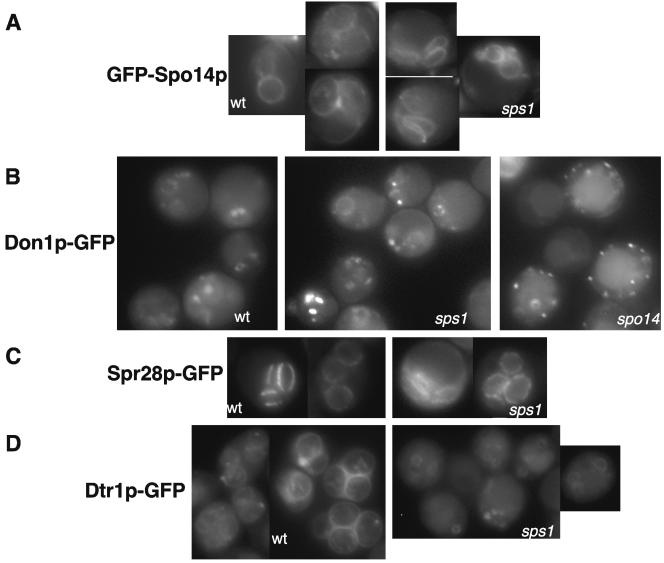
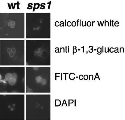
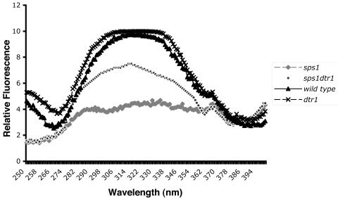
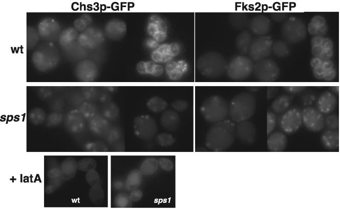
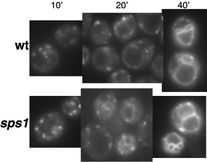
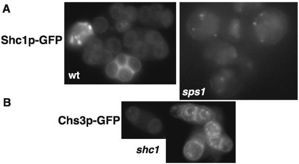
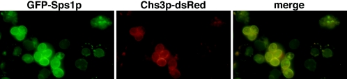
SPS1 encodes a sporulation-specific protein with homology to the Ste20/p21-activated kinase family. Deletion of SPS1 impinges on the formation of the spore wall, which surrounds each of the haploid nuclei generated by the meiotic divisions. Here, we demonstrate that the new internal membranes that surround the meiotic nuclei appear normal in the absence of Sps1p. Analyses of spore wall layers by immunohistochemistry suggest that the inner layers are not efficiently deposited. The defect in spore wall morphogenesis is most likely a consequence of mislocalization of enzymes required for the synthesis of the spore wall layers as both Chs3p, the major chitin synthase in yeast, and Gsc2/Fks2p, a glucan synthase transcriptionally upregulated during sporulation, fail to reach the prospore membrane in the sps1 mutant. Furthermore, localization of Chs3p to the prospore membrane is not dependent on Shc1p, a sporulation-specific homolog of Chs4p, which is required for recruitment of Chs3p to the bud neck in vegetative cells. Sps1p colocalized with Chs3p to peripheral and internal punctate structures and prospore membranes. We propose that Sps1p promotes sporulation, in part, by regulating the intracellular movement of proteins required for spore wall formation.


Figures

FIG. 1.
FIG. 1.
Prospore membrane formation appears normal in sps1 mutants. (A) GFP-Spo14p staining in the wild type (wt) and sps1 mutants; (B) Don1p-GFP staining in the wt and sps1 and spo14Δ mutants. (C) Spr28p-GFP staining in the wt and sps1 mutants; (D) Dtr1p-GFP staining in the wt and sps1 mutants. Living cells were visualized at the time of the meiotic divisions.
FIG. 2.
FIG. 2.
sps1 mutants do not display the full array of spore wall layers. The left panels show wild-type (wt) cells with characteristic staining patterns of calcofluor white (chitosan), anti-glucan antibodies (β-1,3-glucan), and FITC-ConA (mannan) around each individual spore. The fourth spore of the tetrad is out of the plane of focus. sps1 mutants do not display staining with calcofluor white or anti-glucan antibodies around each spore-like structure but do have distinct FITC-ConA staining.
FIG. 3.
FIG. 3.
Dityrosine is synthesized but not incorporated into the spore wall in sps1 mutants. Extracts were prepared and fluorescence was measured as described in Materials and Methods. The intensity of emission was measured at 420 nm. The peak at 315 nm corresponds to dityrosine.
FIG. 4.
FIG. 4.
Chs3p-GFP and Gsc2p-GFP fail to reach the prospore membrane in sps1 mutants. Chs3p-GFP localization is shown in living wild-type (wt) and sps1 cells and with the addition of latA (+latA). Gsc2p-GFP (Fks2p-GFP) localization is shown in living wt and sps1 cells.
FIG. 5.
FIG. 5.
sps1 mutants are proficient for endocytosis. Wild-type (wt) cells and sps1 mutants stained with FM4-64 are shown. Numbers above the panels refer to time (in minutes) after FM4-64 was washed out.
FIG. 6.
FIG. 6.
Shc1p-GFP is not properly localized in sps1 mutants but is not required for Chs3p-GFP localization to the prospore membrane. (A) Shc1p-GFP localization in wild-type (wt) and sps1 cells; (B) Chs3p-GFP localization in shc1Δ cells.
FIG. 7.
FIG. 7.
GFP-Sps1p localizes to internal structures and prospores and displays colocalization with Chs3p-dsRed. Left (green), GFP-Sps1p localization in living cells induced in sporulation; center (red), Chs3p-dsRed localization in living cells induced in sporulation; right (yellow), merged image showing colocalization of GFP-Sps1p and Chs3p-dsRed.

References

    1. Bajgier, B. K., M. Malzone, M. Nickas, and A. M. Neiman. 2001. SPO21 is required for meiosis-specific modification of the spindle pole body in yeast. Mol. Biol. Cell 12:1611-1621. - PMC - PubMed
    1. Briza, P., M. Eckerstorfer, and M. Breitenbach. 1994. The sporulation-specific enzymes encoded by the DIT1 and DIT2 genes catalyze a two-step reaction leading to a soluble LL-dityrosine-containing precursor of the yeast spore wall. Proc. Natl. Acad. Sci. USA 91:4524-4528. - PMC - PubMed
    1. Castillo, A. R., J. B. Meehl, G. Morgan, A. Schutz-Geschwender, and M. Winey. 2002. The yeast protein kinase Mps1p is required for assembly of the integral spindle pole body component Spc42p. J. Cell Biol. 156:453-465. - PMC - PubMed
    1. Christodoulidou, A., V. Bouriotis, and G. Thireos. 1996. Two sporulation-specific chitin deacetylase-encoding genes are required for the ascospore wall rigidity of Saccharomyces cerevisiae. J. Biol. Chem. 271:31420-31425. - PubMed
    1. Chu, S., J. DeRisi, M. Eisen, J. Mulholland, D. Botstein, P. O. Brown, and I. Herskowitz. 1998. The transcriptional program of sporulation in budding yeast. Science 282:699-705. - PubMed

Publication types

MeSH terms