. 2005 Mar;4(3):588-603.
doi: 10.1128/EC.4.3.588-603.2005.

Regulation of the Cdc42/Cdc24 GTPase module during Candida albicans hyphal growth

Affiliations

Regulation of the Cdc42/Cdc24 GTPase module during Candida albicans hyphal growth

Martine Bassilana et al. Eukaryot Cell. 2005 Mar.

Abstract

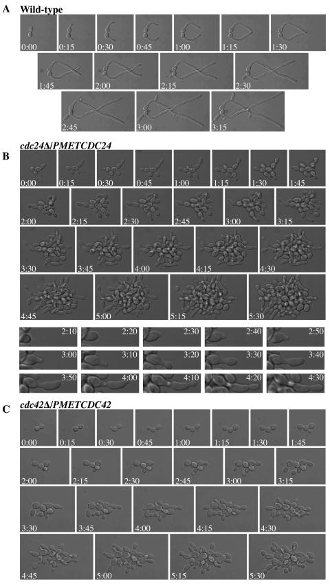
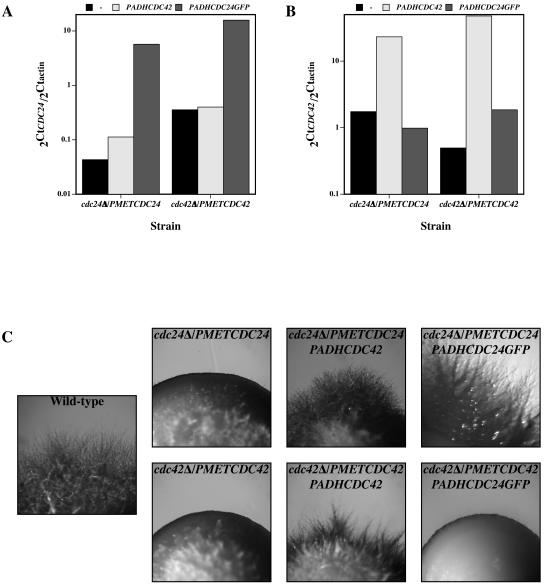
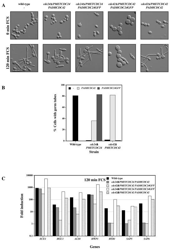
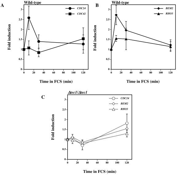
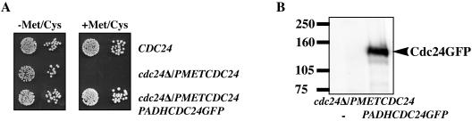
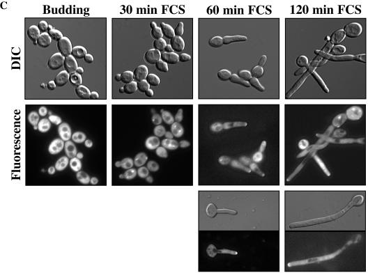
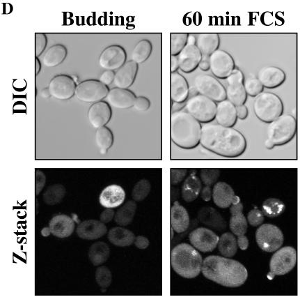
The Rho G protein Cdc42 and its exchange factor Cdc24 are required for hyphal growth of the human fungal pathogen Candida albicans. Previously, we reported that strains ectopically expressing Cdc24 or Cdc42 are unable to form hyphae in response to serum. Here we investigated the role of these two proteins in hyphal growth, using quantitative real-time PCR to measure induction of hypha-specific genes together with time lapse microscopy. Expression of the hypha-specific genes examined depends on the cyclic AMP-dependent protein kinase A pathway culminating in the Efg1 and Tec1 transcription factors. We show that strains with reduced levels of CDC24 or CDC42 transcripts induce hypha-specific genes yet cannot maintain their expression in response to serum. Furthermore, in serum these mutants form elongated buds compared to the wild type and mutant budding cells, as observed by time lapse microscopy. Using Cdc24 fused to green fluorescent protein, we also show that Cdc24 is recruited to and persists at the germ tube tip during hyphal growth. Altogether these data demonstrate that the Cdc24/Cdc42 GTPase module is required for maintenance of hyphal growth. In addition, overexpression studies indicate that specific levels of Cdc24 and Cdc42 are important for invasive hyphal growth. In response to serum, CDC24 transcript levels increase transiently in a Tec1-dependent fashion, as do the G-protein RHO3 and the Rho1 GTPase activating protein BEM2 transcript levels. These results suggest that a positive feedback loop between Cdc24 and Tec1 contributes to an increase in active Cdc42 at the tip of the germ tube which is important for hypha formation.


Figures

FIG.1.
FIG.1.
Cdc24 and Cdc42 levels are reduced in cdc24Δ/PMETCDC24 and cdc42Δ/PMETCDC42 strains. (A) Transcript levels of CDC24 and CDC42 in the wild type and ectopic expression mutants. RNA transcript levels normalized to actin transcript levels (2Ctgene/2Ctactin) were determined from budding cells grown at 30°C in YEPD-Uri medium. Values are the means of three or four independent experiments, with bars representing standard deviations. (B) Cdc42 protein levels in the wild type and ectopic expression mutants. Immunoblot analysis of Cdc42 protein levels from the indicated strains grown in YEPD-Uri medium at 30°C. Anti-Cdc42 polyclonal antibodies were used. The upper band is a nonspecific protein cross-reacting with anti-Cdc42 antibody.
FIG. 2.
FIG. 2.
Quantitative analyses of hyphal gene and transcription factor expression in wild-type cells exposed to FCS. (A) Normalized transcript levels of HSGs in wild-type cells responding to FCS at 37°C. Relative transcript abundance values (2Ctgene/2Ctactin) are the means of three independent experiments with standard deviations indicated. (B) Normalized transcript levels of transcription factors in wild-type cells responding to FCS at 37°C. Relative transcript abundance values (2Ctgene/2Ctactin) are the means of three independent experiments with standard deviations indicated. (C) Representative images of cells responding to FCS. DIC images of fixed cells from the same experiment were recorded at the indicated times.
FIG. 3.
FIG. 3.
In response to FCS cdc24Δ/PMETCDC24 and cdc42Δ/PMETCDC42 mutants induce HSGs yet are unable to maintain induced levels. Fold induction of HSGs after incubation of cells for 10 (A), 30 (B), and 120 (C) min in FCS at 37°C is shown. Fold induction is the normalized transcript level at the indicated time divided by the normalized transcript level at time zero. The mean values of two independent experiments are shown. (D) Representative images of cells responding to FCS. DIC images of fixed cells from the same experiment were recorded at the indicated times.
FIG. 4.
FIG. 4.
cdc24Δ/PMETCDC24 and cdc42Δ/PMETCDC42 mutants initiate a morphological response to serum but are unable to form hyphae. (A) Time lapse microscopy of wild-type cells in FCS at 37°C. DIC images were captured every 5 min over 6 h, and images every 15 min are shown over 195 min. (B) Time lapse microscopy of cdc24Δ/PMETCDC24 mutant cells in FCS at 37°C. DIC images were captured every 5 min over 6 h, and images every 15 min are shown during 330 min (top panels). Close-ups of individual cell images (bottom panels) every 10 min from 130 to 270 min are also shown. (C) Time lapse microscopy of cdc42Δ/PMETCDC42 mutant cells in FCS at 37°C. DIC images were captured every 5 min over 6 h, and images every 15 min are shown during 330 min.
FIG. 5.
FIG. 5.
Overexpression of CDC42 in a cdc24Δ/PMETCDC24 mutant partially suppresses the invasive growth defect. Normalized transcript levels of CDC24 (A) and CDC42 (B) in indicated strains overexpressing CDC42 and CDC24GFP are shown. Relative transcript abundance values (2Ctgene/2Ctactin) are the means of two independent experiments and were determined as for Fig. 3C. (C) Invasive growth of indicated strains overexpressing CDC42 or CDC24GFP. Images were taken after 8 days growth on YEPD-Uri-FCS agar at 37°C.
FIG. 6.
FIG. 6.
Overexpression of CDC42 in a cdc24Δ/PMETCDC24 mutant partially restores germ tube formation and HSG induction. (A) Representative images of cells responding to FCS. DIC images of fixed cells were recorded at the indicated times. (B) Percentage of cells with germ tubes in indicated strains after 120 min in FCS at 37°C. Values (n = 200 to 250 cells) are from the experiment shown in panel A, with similar results observed in three independent experiments. (C) Fold induction of HSGs after 120 min in FCS at 37°C. Fold induction, determined as for Fig. 3C, of transcript levels from the same experiment for which results are shown in panels A and B.
FIG. 7.
FIG. 7.
In response to FCS, CDC24 transcript levels transiently increase in a Tec1-dependent fashion. (A) Levels of CDC24 and CDC42 transcripts in wild-type cells incubated with FCS at 37°C. Fold induction was determined as for Fig. 3C, and values are the averages of five independent experiments, with the bars indicating standard deviations. (B) Levels of BEM2 and RHO3 transcripts in wild-type cells incubated with FCS at 37°C. Fold induction was determined as above, and values are the averages of five independent experiments, with bars indicating standard deviations. (C) Levels of CDC24, BEM2, and RHO3 transcripts in tec1Δ/tec1Δ cells incubated with FCS at 37°C. Fold induction was determined as above, and values are the averages of three independent experiments, with bars indicating standard deviations.
FIG. 8.
FIG. 8.
Cdc24 is recruited to the tips of germ tubes in response to FCS. (A) Cdc24GFP is functional. The indicated strains were grown on medium which permits (−Met/Cys) or represses (+Met/Cys) MET3-driven CDC24 expression for 2 days at 30°C. (B) Immunoblot analysis of cdc24Δ/PMETCDC24 mutant cells with or without integrated Cdc24GFP. Extracts from cells grown at 30°C were analyzed with anti-GFP polyclonal antibodies. (C) Cdc24GFP localization over 120 min in cells in the presence of FCS. cdc24Δ/PMETCDC24 cells overexpressing Cdc24GFP were grown in YEPD-Uri medium with Met and Cys at 30°C (Budding) and for the indicated times in FCS at 37°C. DIC and fluorescence images are shown. Panels show individual cells after 60 and 120 min in FCS at 37°C. (D) Cdc24GFP localization in the presence or absence of FCS. cdc24Δ/PMETCDC24 cells overexpressing Cdc24GFP grown in SC medium with Met and Cys for 60 min in the presence or absence of FCS were examined by confocal microscopy. DIC images are average intensity projections, and fluorescence images are maximum intensity projections of 15 to 17 0.2-μm z sections.
FIG. 8.
FIG. 8.
Cdc24 is recruited to the tips of germ tubes in response to FCS. (A) Cdc24GFP is functional. The indicated strains were grown on medium which permits (−Met/Cys) or represses (+Met/Cys) MET3-driven CDC24 expression for 2 days at 30°C. (B) Immunoblot analysis of cdc24Δ/PMETCDC24 mutant cells with or without integrated Cdc24GFP. Extracts from cells grown at 30°C were analyzed with anti-GFP polyclonal antibodies. (C) Cdc24GFP localization over 120 min in cells in the presence of FCS. cdc24Δ/PMETCDC24 cells overexpressing Cdc24GFP were grown in YEPD-Uri medium with Met and Cys at 30°C (Budding) and for the indicated times in FCS at 37°C. DIC and fluorescence images are shown. Panels show individual cells after 60 and 120 min in FCS at 37°C. (D) Cdc24GFP localization in the presence or absence of FCS. cdc24Δ/PMETCDC24 cells overexpressing Cdc24GFP grown in SC medium with Met and Cys for 60 min in the presence or absence of FCS were examined by confocal microscopy. DIC images are average intensity projections, and fluorescence images are maximum intensity projections of 15 to 17 0.2-μm z sections.

References

    1. Akashi, T., T. Kanbe, and K. Tanaka. 1994. The role of the cytoskeleton in the polarized growth of the germ tube in Candida albicans. Microbiology 140:271-280. - PubMed
    1. Anderson, J. M., and D. R. Soll. 1986. Differences in actin localization during bud and hypha formation in the yeast Candida albicans. J. Gen. Microbiol. 132:2035-2047. - PubMed
    1. Arkowitz, R. A., and N. Lowe. 1997. A small conserved domain in the yeast Spa2p is necessary and sufficient for its polarized localization. J. Cell Biol. 138:17-36. - PMC - PubMed
    1. Ayscough, K. R., and D. G. Drubin. 1998. A role for the yeast actin cytoskeleton in pheromone receptor clustering and signalling. Curr. Biol. 8:927-930. - PubMed
    1. Bailey, D. A., P. J. Feldmann, M. Bovey, N. A. Gow, and A. J. Brown. 1996. The Candida albicans HYR1 gene, which is activated in response to hyphal development, belongs to a gene family encoding yeast cell wall proteins. J. Bacteriol. 178:5353-5360. - PMC - PubMed

Publication types

MeSH terms

LinkOut - more resources