. 2005 Apr;3(4):e87.
doi: 10.1371/journal.pbio.0030087. Epub 2005 Mar 15.

Adult murine skeletal muscle contains cells that can differentiate into beating cardiomyocytes in vitro

Affiliations

Adult murine skeletal muscle contains cells that can differentiate into beating cardiomyocytes in vitro

Steve O Winitsky et al. PLoS Biol. 2005 Apr.

Abstract

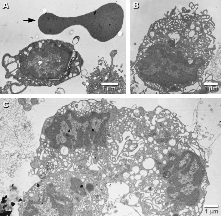
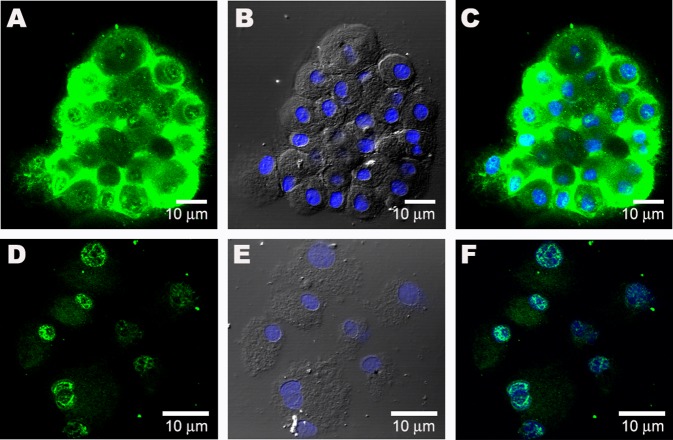
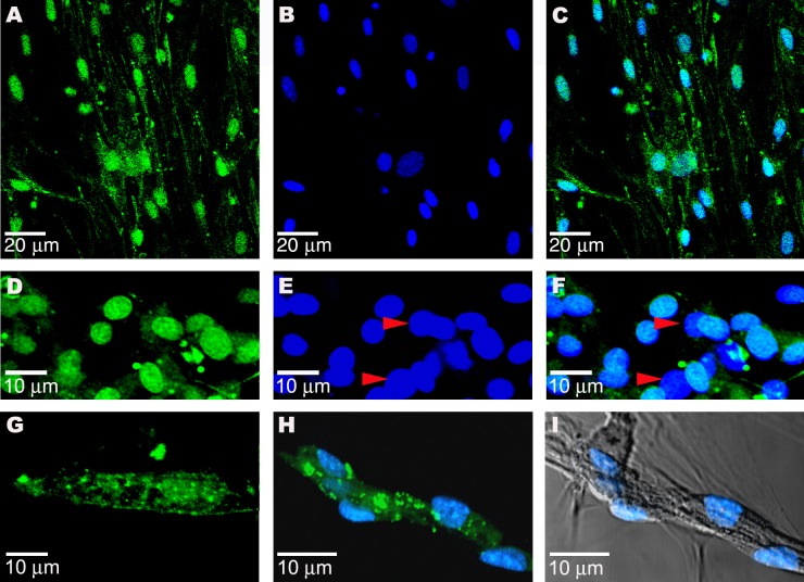
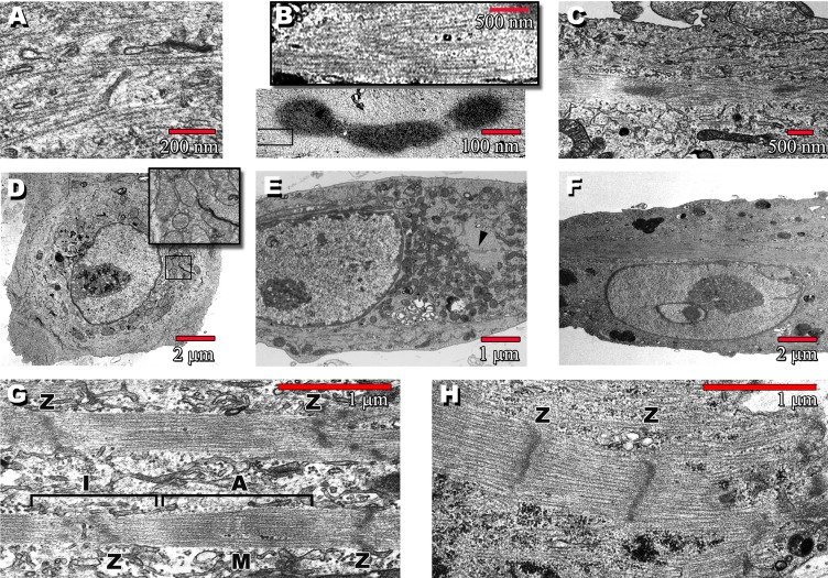
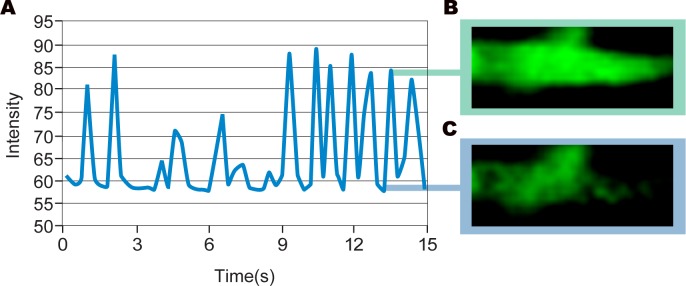
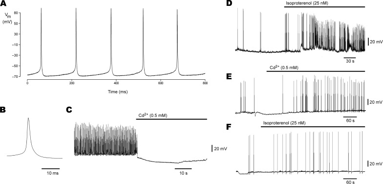
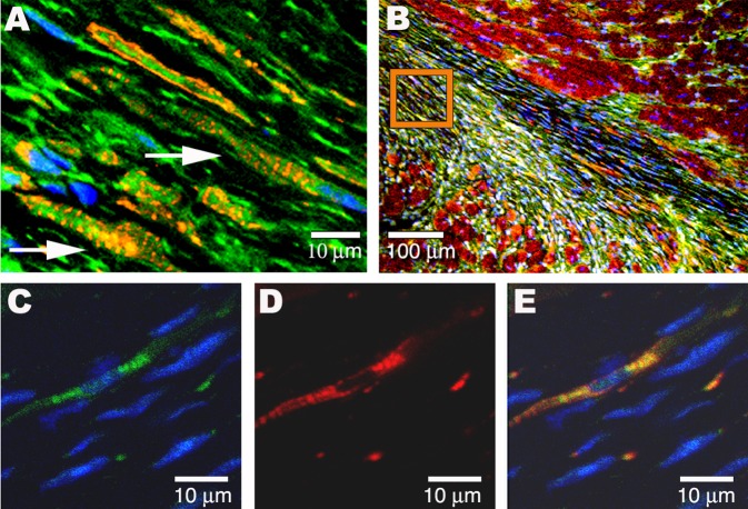
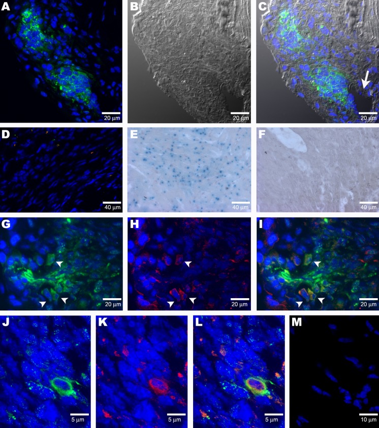
It has long been held as scientific fact that soon after birth, cardiomyocytes cease dividing, thus explaining the limited restoration of cardiac function after a heart attack. Recent demonstrations of cardiac myocyte differentiation observed in vitro or after in vivo transplantation of adult stem cells from blood, fat, skeletal muscle, or heart have challenged this view. Analysis of these studies has been complicated by the large disparity in the magnitude of effects seen by different groups and obscured by the recently appreciated process of in vivo stem-cell fusion. We now show a novel population of nonsatellite cells in adult murine skeletal muscle that progress under standard primary cell-culture conditions to autonomously beating cardiomyocytes. Their differentiation into beating cardiomyocytes is characterized here by video microscopy, confocal-detected calcium transients, electron microscopy, immunofluorescent cardiac-specific markers, and single-cell patch recordings of cardiac action potentials. Within 2 d after tail-vein injection of these marked cells into a mouse model of acute infarction, the marked cells are visible in the heart. By 6 d they begin to differentiate without fusing to recipient cardiac cells. Three months later, the tagged cells are visible as striated heart muscle restricted to the region of the cardiac infarct.


Figures

Figure 1
Figure 1. EMs of Day 0 Spoc Cells
Electron micrograph of a freshly isolated Spoc cell (A) shows its small size relative to the typical biconcave profile of the red blood cell above it (arrow). (B) shows a typical Spoc cell with a large number of vesicular structures, lamellipodia, and filopodia. (C) displays three Spoc cells so tightly clumped that their borders are difficult to distinguish. This typical clumping makes fluorescence-activated cell sorting (FACS) analysis difficult.
Figure 2
Figure 2. Sublocalization of GATA-4 in Spoc Cells
(A) GATA-4 is detected in the cytoplasm of day 10 Spoc cells (cytospin). (B) Nomarski image of (A), showing DAPI-stained blue nuclei. (C) Merge image showing nuclei and GATA-4 staining. (D) When Spoc cells are incubated with 20 μM isoproterenol for 1 h, the GATA-4 nuclear staining is seen. (E) Nomarski image of (D). (F) Merge of (D) and (E), showing sublocalization of GATA-4 to nuclei. A weaker GATA-4 signal is present in the cytoplasm.
Figure 3
Figure 3. CPS Cells Stain Positive for Cardiac-Specific Proteins
(A) GATA-4 in day 7 CPS cells. (B) Nuclear staining with DAPI. (C) Overlay of (A) and (B). (D) Nkx-2.5 is detected in the nuclei of round, day 21 beating cells (green). (E) Noncardiac cells (red arrowheads) do not show nuclear staining for Nkx-2.5. (F) Overlay of (D) and (E). (G) Beating cells, after 28 d in culture, stain positive for cardiac L-type Ca++ channel. (H) Connexin 43 (green) in cluster of uninucleate day 21 beating cells in culture. (I) Nomarski light micrograph (differential interference contrast) of cell cluster in (H).
Figure 4
Figure 4. Transmission EMs Show the Post-Replating Progression of CPS Cells
(A) Round, day 3 cells contain disordered myosin filaments. Some of these cells beat while still floating (see Video S1) and typically have APs as shown in Figure 6A. (B) Upper box is a blowup taken from lower panel, showing myosin filaments of characteristic 1.6-μm length radiating outward from dense body. (C) Day 14 cell with a single, central nucleus shows a stretching out of the dense bodies into an organizing sarcomere. (D) Day 3 round cells containing copious mitochondria (inset). (E) Elongated day 7 cell containing a dense body (arrowhead). (F) Uninucleate day 14 cell, same cell as in (C). (G) By day 56, a well-defined sarcomere is present, with identifiable A- and I-bands and M- and Z-lines. (H) Sarcomere from a fetal cardiomyocyte is shown for comparison.
Figure 5
Figure 5. Measuring Calcium Transient Frequency
Graphical representation of the calcium transient in a beating CPS cell–derived cardiomyocyte (A). Fluorescent intensity is proportional to the amount of calcium binding to fluo-3 dye upon release of calcium from the sarcoplasmic reticulum. Peak intensity (B) and baseline (C) are shown.
Figure 6
Figure 6. Whole-Cell Voltage Recordings from Spoc Cell–Derived Cardiomyocytes
(A) Spontaneous AP firing in a nonbeating, teardrop-shaped cell. (B) Representative AP from recording in (A) on an expanded time scale; AP threshold is –60 mV. (C) Action potential firing in another cell is blocked upon bath perfusion with 0.5 mM cadmium chloride (horizontal bar). (D) Acceleration of AP firing upon perfusion with 25 nM isoproterenol (horizontal bar) is demonstrated, indicating the presence of adrenergic receptors on these cells. (E) Skeletal myotube APs, if present, differ in that their frequency is unaffected by Cd++. (F) Isoproterenol also does not affect skeletal muscle AP frequency.
Figure 7
Figure 7. In Vivo Myocardial Infarction Transplantation Studies
(A) The GFP-tagged Spoc cells (green), unfractionated for Sca-1, injected into the peripheral blood of a murine acute infarct model have developed after 14 wk into cardiomyocytes (arrows) within the infarct region. Donor Spoc cell–derived GFP cardiomyocytes are characterized by single central nuclei and striations staining for RLCP (red). (B) Longitudinal fresh-frozen tissue slice showing the region of infarct from which (A) was taken (orange box) and adjacent normal endogenous cardiac tissue (RLCP, red). (C) GFP+/Sca-1 Spoc cells were injected into the peripheral circulation of a murine acute MI model and are detected in the infarct after 4 wk by expression of GFP (green). (D) RLCP (red) is also expressed. (E) Overlay of (C) and (D).
Figure 8
Figure 8. Cre Expressor/Beta-Galactosidase Reporter Myocardial Infarction Studies
(A) Nests of Cre+ cells (green) are detected 1 wk after tail-vein injection into an acute infarct model. (B) Nomarski image of (A). (C) Merged image of (A) and (B). The clusters are located near a blood vessel (arrow). (D) Infarcted tissue in a control MI model (infarction surgery but no donor-cell injection) showing a lack of staining for Cre (no green) and GATA-4 (no red). (E) Control X-gal staining of ROSA mouse heart. (F) In a sequential series of tissue sections, odd-numbered sections were immunostained for Cre, yielding the results seen in (A). These two clusters of cells were seen on five sections (sections 1, 3, 5, 7, and 9). Even-numbered sections (sections 2, 4, 6, and 8) were stained for X-gal. No X-gal+ cells were found. One slide was immunostained, showing the Cre+ cells present. This slide was then stained for X-gal and was found to be X-gal. The lack of X-gal staining of the serial sections indicates that at 1 wk no fusion of donor and host cells has occurred in the infarct. (G) Cluster of Cre+ donor cells detected in infarcted heart tissue of a 1-wk-old acute infarct model. Arrowheads indicate cells that also express GATA-4, as shown in (H). (H) GATA-4 (red) is mostly present in some cells in the margin of the cluster. Arrowheads indicate cells that also express Cre, as shown in (G) (I) Merged image showing co-localization (arrowheads) of Cre (green) with GATA-4 (red) in some cells of the cluster of Cre+ cells. (J–L) Co-staining of donor cells with anti–Cre antibody (green) and MSC 21 (red) is apparent after 7 d in an acute infarct model. (M) There is a lack of staining with MSC 21 (no red) in the infracted tissue of mice that have not received Spoc cell injections.

Comment in

References

    1. Condorelli G, Borello U, De Angelis L, Latronico M, Sirabella D, et al. Cardiomyocytes induce endothelial cells to trans-differentiate into cardiac muscle: Implications for myocardium regeneration. Proc Natl Acad Sci U S A. 2001;98:10733–10738. - PMC - PubMed
    1. Kehat I, Kenyagin-Karsenti D, Snir M, Segev H, Amit M, et al. Human embryonic stem cells can differentiate into myocytes with structural and functional properties of cardiomyocytes. J Clin Invest. 2001;108:407–414. - PMC - PubMed
    1. Leor J, Patterson M, Quinones MJ, Kedes LH, Kloner RA. Transplantation of fetal myocardial tissue into the infarcted myocardium of rat. A potential method for repair of infarcted myocardium? Circulation. 1996;94:II332–II336. - PubMed
    1. Bjornson CR, Rietze RL, Reynolds BA, Magli MC, Vescovi AL. Turning brain into blood: A hematopoietic fate adopted by adult neural stem cells in vivo. Science. 1999;283:534–537. - PubMed
    1. Deasy BM, Jankowski RJ, Huard J. Muscle-derived stem cells: Characterization and potential for cell-mediated therapy. Blood Cells Mol Dis. 2001;27:924–933. - PubMed