. 2005 May;16(5):2414-23.
doi: 10.1091/mbc.e04-10-0883. Epub 2005 Mar 9.

Initiation of embryonic cardiac pacemaker activity by inositol 1,4,5-trisphosphate-dependent calcium signaling

Affiliations

Initiation of embryonic cardiac pacemaker activity by inositol 1,4,5-trisphosphate-dependent calcium signaling

Annabelle Méry et al. Mol Biol Cell. 2005 May.

Abstract

In the adult, the heart rate is driven by spontaneous and repetitive depolarizations of pacemaker cells to generate a firing of action potentials propagating along the conduction system and spreading into the ventricles. In the early embryo before E9.5, the pacemaker ionic channel responsible for the spontaneous depolarization of cells is not yet functional. Thus the mechanisms that initiate early heart rhythm during cardiogenesis are puzzling. In the absence of a functional pacemaker ionic channel, the oscillatory nature of inositol 1,4,5-trisphosphate (InsP3)-induced intracellular Ca2+ signaling could provide an alternative pacemaking mechanism. To test this hypothesis, we have engineered pacemaker cells from embryonic stem (ES) cells, a model that faithfully recapitulates early stages of heart development. We show that InsP3-dependent shuttle of free Ca2+ in and out of the endoplasmic reticulum is essential for a proper generation of pacemaker activity during early cardiogenesis and fetal life.


Figures

Figure 1.
Figure 1.
Cells expressing EYFP under control of the α-MHC promoter are true pacemaker cells. (A) α-MHC expression was measured by real-time quantitative PCR after reverse transcription of mRNA extracted from embryonic ventricles and atria at different stages of development (E7, E9, E14.5, E17.5) or after birth in neonatal mice (Neo). Data are normalized to β-tubulin expression and expressed as fold increase in expression compared with E7 embryonic heart α-MHC mRNA content. CGR8 ES cell clones expressing EYFP under control of the α-MHC promoter (B) or of the cardiac α-actin promoter (C) were allowed to differentiate into cardiomyocytes within embryoid bodies (EBs). EYFP was visualized by confocal microscopy at 10× (BC) and 63× magnification (inset B). (D) Sarcomeric units of differentiating myocytes including some expressing EYFP under control of the α-MHC promoter were immunostained within EBs with an anti-actinin antibody and anti-mouse secondary antibody coupled to Alexa350 and visualized by confocal microscopy (63× magnification). (E) Cardiomyocytes including α-MHC-EYFP–expressing cells were immunostained with an anti-β-MHC antibody and an anti-mouse secondary antibody coupled to Alexa546 and imaged by confocal microscopy (63× magnification). (F) Myocytes expressing EYFP under control of the α-MHC promoter were microdissected out of day 8–10 EBs. RNA was extracted from the cells; EYFP, Cx45, and HCN4 were amplified by real-time quantitative PCR. Profiles of melting curves obtained at the end of the PCR run are illustrated; the amplified fragments were further separated on agarose gel to identify the products (inset). (G) Other EYFP-expressing cells were enzymatically dissociated using collagenase and cultured for 24–48 h. Action potentials (G) and the If current (H) were recorded in these cells under microelectrode or voltage-clamp configuration, respectively. The figure is representative of 6–8 experiments. *Significantly different from D9 (p ≤ 0.05)
Figure 2.
Figure 2.
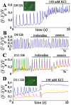
Pacemaker activity of day 9 embryoid bodies (EBs) does not depend upon a voltage-sensitive ionic conductance. Fluo-4–loaded day 9 EBs were challenged with 140 mM KCl (KCl replaced NaCl) (A) or with 10 μM ivabradine or 1 μM zeneca (B). Fluo-4–loaded day 11 EBs were challenged with 10 μM ivabradine or 1 μM zeneca (C) or with 140 mM KCl (D). Recordings show Ca2+ spikes within selected regions of interest (ROI) and are expressed as ΔF/Fo, where Fo is the lowest basal fluorescence recorded at the beginning of the experiment. The figure is representative of at least five experiments.
Figure 3.
Figure 3.
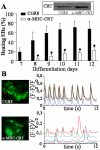
Overexpression of calreticulin in ES cell–derived pacemaker cells impairs excitability and contractility of embryoid bodies (EBs). (A) Wild-type CGR8 ES cells or cells expressing calreticulin under control of the α-MHC promoter were allowed to differentiate within EBs and beating activity of EBs was monitored daily from day 7. The figure is a mean of three separate differentiation experiments. The inset shows an anti-calreticulin Western blot of proteins extracted from wild-type CGR8 or α-MHC-calreticulin EBs. Wild-type CGR8 or α-MHC-calreticulin EBs were loaded with fluo-4 and Ca2+ transients were monitored in ES cell–derived cardiomyocytes using fast acquisition confocal microscopy (B). Recordings show Ca2+ spikes within selected regions of interest (ROI) and are expressed as ΔF/Fo, where Fo is the lowest basal fluorescence recorded at the beginning of the experiment. The figure is representative of at least five experiments.
Figure 4.
Figure 4.
InsP3RIs are required to initiate pacemaker activity in embryoid bodies (EBs). (A) Two clones of ES cells, IP3Ras13 and IP3Ras14, were selected for their level of expression of the InsP3RI antisense cDNA. Left panel, an agarose gel electrophoresis of real time quantitative PCR products amplified after reverse transcription of the antisense mRNA from total RNA of day 12 EB. Wild-type CGR8 d 12 EBs (CGR8) and no DNA in PCR reaction (-) served as negative controls. The α-MHC-InsP3RI antisense plasmid (+) served as a positive control. Right panel, two embryonic cardiomyocytes cotransfected with a plasmid (α-MHC-InsP3RI) encoding the InsP3RI antisense together with a plasmid encoding a fusion protein myosin light chain 2v-GFP (MLC2vGFP) to track the transfected cells and their neighbor nontransfected cardiomyocyte. Cells were stained 72 h after transfection with an anti-InsP3RI antibody. The white arrows show the absence of InsP3RIs in the nuclear area of the antisense-transfected cells (n = 2 experiments, 55 scored cells), whereas the yellow arrow shows the InsP3RIs located in the nuclear envelope and emanating ER of the nontransfected cell. (B) ES cells expressing the InsP3RI antisense cDNA under control of the α-MHC promoter were allowed to differentiate within EBs and beating activity of EBs was monitored daily from day 7. The figure is a mean of two separate differentiation experiments. (C) Day 9–10 CGR8 or IP3Ras EBs were loaded with fluo-4 and Ca2+ transients were monitored in ES cell–derived cardiomyocytes using fast acquisition confocal microscopy. (D) Xestospongin (5 μM) was applied to wild-type fluo-4–loaded EBs. Right panel, Ca2+ spiking was recorded after 2-min exposure to the inhibitor; in the same running experiments, Ca2+ spikes were fully abolished after 5 min superfusion of the drug. Recordings show Ca2+ spikes within selected regions of interest (ROIs) and are expressed as ΔF/Fo, where Fo is the lowest basal fluorescence recorded at the beginning of the experiment. The figure is representative of 3–5 separate experiments. (E) Xestospongin (5 μM) was applied to E9–9.5 mouse embryonic heart loaded with fluo-4 and observed at 10× magnification. Ca2+ spiking was recorded after 2-min exposure to the inhibitor; in the same running experiments, Ca2+ spikes were fully abolished after 5-min superfusion of the drug. Recordings show Ca2+ spikes within selected regions of interest (ROIs) located in the atrium (a) or ventricle (v) of the heart tube and are expressed as ΔF/Fo, where Fo is the lowest basal fluorescence recorded at the beginning of the experiment. The figure is representative of three separate experiments.
Figure 5.
Figure 5.
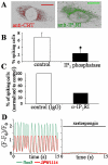
InsP3-induced Ca2+ mobilization regulates pacemaker activity of E14.5 embryonic pacemaker atrial cells. (A) Cells were isolated from atria of E14.5 embryos and cultured for 24–48 h. ER of embryonic cardiomyocytes was immunostained with anticalreticulin and anti-InsP3RI antibodies and visualized by confocal microscopy. Scale bar, 10 μm. (B) Cells were transfected with a vector encoding Ds Red alone or with both Ds Red and a pcDNA vector encoding InsP3 phosphatase. After loading with fluo-4, Ca2+ transients were recorded in isolated cells. The figure is a mean of three separate experiments including 507 cells expressing Ds Red only and 511 cells expressing both Ds Red and the InsP3 phosphatase. (C) Cells were microinjected with micropipettes containing 2.5 mM fluo-3 and 500 μg/ml 18A10 monoclonal anti-InsP3RI antibody or mouse IgGs. Ca2+ transients were recorded in 37 isolated cells microinjected with control IgGs and 47 isolated cells microinjected with the 18A10 antibody. (D) Cells were microinjected with micropipettes containing 2.5 mM fluo-3 and 3 mg/ml JPW1114. Ca2+ transients, and action potential were simultaneously recorded in isolated cells before and after 2 or 5 min of xestospongin application (5 μM). The figure is representative of six experiments.
Figure 6.
Figure 6.
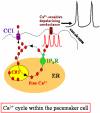
Model of InsP3-induced spontaneous activity in embryonic pacemaker cells. Ca2+ released upon InsP3 binding to its receptor activates a depolarizing conductance (Na+/Ca2+ exchanger and/or a nonspecific cationic and depolarizing conductance) and thus triggers action potential firing. CCI allows for refilling the ER. This Ca2+ cycling acting in a rhythmic and perpetual manner is a predominant pacemaking mechanism in early cardiogenesis and regulates or is required together with the If current as a basis of fetal pacemaking mechanism.

Similar articles

Cited by

References

    1. Accili, E. A., Proenza, C., Baruscotti, M., and DiFrancesco, D. (2002). From funny current to HCN channels: 20 years of excitation. News Physiol. Sci. 17, 32-37. - PubMed
    1. Behfar, A., Zingman, L., Hodgson, D., Rauzier, J., Kane, G., Terzic, A., and Pucéat, M. (2002). Stem cell differentiation requires a paracrine pathway in the heart. FASEB J. 16, 1558-1566. - PubMed
    1. Berridge, M. J., Bootman, M. D., and Roderick, H. L. (2003). Calcium signalling: dynamics, homeostasis and remodelling. Nat. Rev. Mol. Cell. Biol. 4, 517-529. - PubMed
    1. Berridge, M. J., Lipp, P., and Bootman, M. D. (2000). The versatility and universality of calcium signalling. Nat. Rev. Mol. Cell. Biol. 1, 11-21. - PubMed
    1. Bony, C., Roche, S., Sasaki, T., Crackowe, M., Penninger, J., Mano, H., and Pucéat, M. (2001). A specific role of phosphatidylinositol 3-kinase γ: a regulation of autonomic Ca2+ oscillations in cardiac cells. J. Cell Biol. 152, 717-728. - PMC - PubMed

Publication types

MeSH terms