

Long-term in vivo provision of antigen-specific T cell immunity by programming hematopoietic stem cells
- PMID: 15758071
- PMCID: PMC553287
- DOI: 10.1073/pnas.0500600102
Long-term in vivo provision of antigen-specific T cell immunity by programming hematopoietic stem cells
Abstract
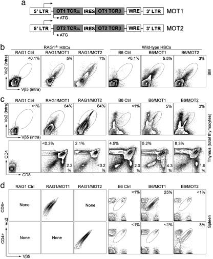
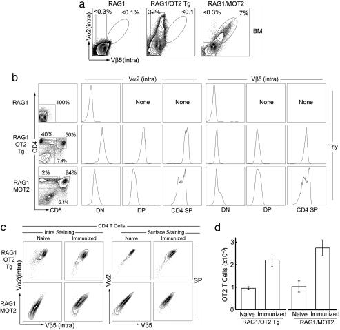
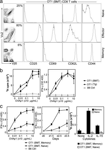
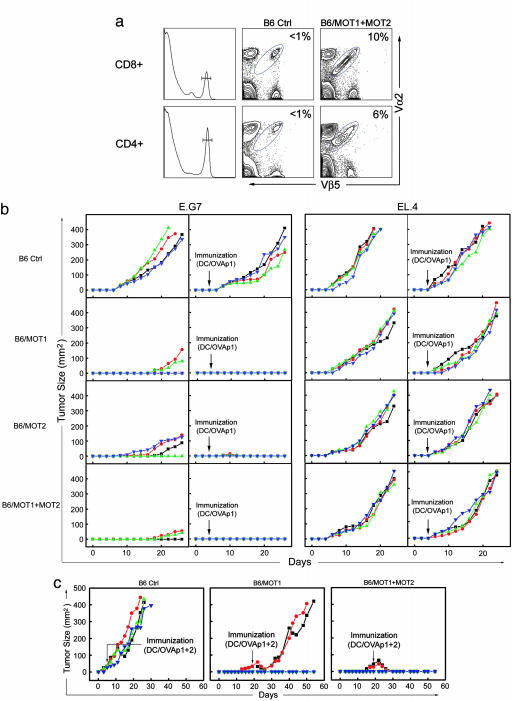
A method to genetically program mouse hematopoietic stem cells to develop into functional CD8 or CD4 T cells of defined specificity in vivo is described. For this purpose, a bicistronic retroviral vector was engineered that efficiently delivers genes for both alpha and beta chains of T cell receptor (TCR) to hematopoietic stem cells. When modified cell populations were used to reconstruct the hematopoietic lineages of recipient mice, significant percentages of antigen-specific CD8 or CD4 T cells were observed. These cells expressed normal surface markers and responded to peptide antigen stimulation by proliferation and cytokine production. Moreover, they could mature into memory cells after peptide stimulation. Using TCRs specific for a model tumor antigen, we found that the recipient mice were able to partially resist a challenge with tumor cells carrying the antigen. By combining cells modified with CD8- and CD4-specific TCRs, and boosting with dendritic cells pulsed with cognate peptides, complete suppression of tumor could be achieved and even tumors that had become established would regress and be eliminated after dendritic cell/peptide immunization. This methodology of "instructive immunotherapy" could be developed for controlling the growth of human tumors and attacking established pathogens.
Figures
Similar articles
-
RAG-dependent peripheral T cell receptor diversification in CD8+ T lymphocytes.Proc Natl Acad Sci U S A. 2002 Nov 26;99(24):15566-71. doi: 10.1073/pnas.242321099. Epub 2002 Nov 13. Proc Natl Acad Sci U S A. 2002. PMID: 12432095 Free PMC article.
-
Transplantation of mouse HSCs genetically modified to express a CD4-restricted TCR results in long-term immunity that destroys tumors and initiates spontaneous autoimmunity.J Clin Invest. 2010 Dec;120(12):4273-88. doi: 10.1172/JCI43274. Epub 2010 Nov 15. J Clin Invest. 2010. PMID: 21084750 Free PMC article.
-
Dual antigen target-based immunotherapy for prostate cancer eliminates the growth of established tumors in mice.Immunotherapy. 2011 Jun;3(6):735-46. doi: 10.2217/imt.11.59. Immunotherapy. 2011. PMID: 21668311
-
Potential use of T cell receptor genes to modify hematopoietic stem cells for the gene therapy of cancer.Pathol Oncol Res. 1999;5(1):3-15. doi: 10.1053/paor.1999.0003. Pathol Oncol Res. 1999. PMID: 10079371 Review.
-
Biological properties of dendritic cells: implications to their use in the treatment of cancer.Mol Med Today. 1997 Jun;3(6):254-60. doi: 10.1016/S1357-4310(97)01047-2. Mol Med Today. 1997. PMID: 9211416 Review.
Cited by
-
Epigenetic Suppression of Transgenic T-cell Receptor Expression via Gamma-Retroviral Vector Methylation in Adoptive Cell Transfer Therapy.Cancer Discov. 2020 Nov;10(11):1645-1653. doi: 10.1158/2159-8290.CD-20-0300. Epub 2020 Jul 22. Cancer Discov. 2020. PMID: 32699033 Free PMC article.
-
Immunogenicity and tumorigenicity of pluripotent stem cells and their derivatives: genetic and epigenetic perspectives.Curr Stem Cell Res Ther. 2014 Jan;9(1):63-72. doi: 10.2174/1574888x113086660068. Curr Stem Cell Res Ther. 2014. PMID: 24160683 Free PMC article. Review.
-
Pseudotyping lentiviral vectors with lymphocytic choriomeningitis virus glycoproteins for transduction of dendritic cells and in vivo immunization.Hum Gene Ther Methods. 2014 Dec;25(6):328-38. doi: 10.1089/hgtb.2014.105. Hum Gene Ther Methods. 2014. PMID: 25416034 Free PMC article.
-
Site-specific labeling of enveloped viruses with quantum dots for single virus tracking.ACS Nano. 2008 Aug;2(8):1553-62. doi: 10.1021/nn8002136. ACS Nano. 2008. PMID: 19079775 Free PMC article.
-
Human cancer-targeted immunity via transgenic hematopoietic stem cell progeny.Nat Commun. 2025 Jul 1;16(1):5599. doi: 10.1038/s41467-025-60816-z. Nat Commun. 2025. PMID: 40593578 Free PMC article. Clinical Trial.
References
-
- Chambers, C. A., Kuhns, M. S., Egen, J. G. & Allison, J. P. (2001) Annu. Rev. Immunol. 19, 565–594. - PubMed
-
- Smyth, M. J., Godfrey, D. I. & Trapani, J. A. (2001) Nat. Immunol. 2, 293–299. - PubMed
-
- Matzinger, P. (2002) Science 296, 301–305. - PubMed
-
- Ochsenbein, A. F. (2002) Cancer Gene. Ther. 9, 1043–1055. - PubMed
-
- Marincola, F. M., Wang, E., Herlyn, M., Seliger, B. & Ferrone, S. (2003) Trends Immunol. 24, 335–342. - PubMed
Publication types
MeSH terms
Substances
Grants and funding
LinkOut - more resources
Full Text Sources
Other Literature Sources
Medical
Research Materials
