. 2005 Mar 22;102(12):4566-71.
doi: 10.1073/pnas.0407357102. Epub 2005 Mar 9.

High-dose radiation with bone marrow transfer prevents neurodegeneration in an inherited glaucoma

Affiliations

High-dose radiation with bone marrow transfer prevents neurodegeneration in an inherited glaucoma

Michael G Anderson et al. Proc Natl Acad Sci U S A. .

Abstract

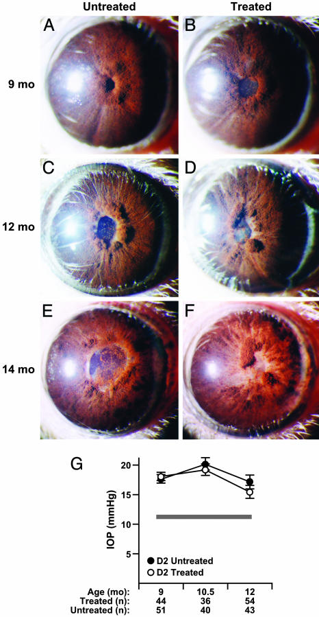
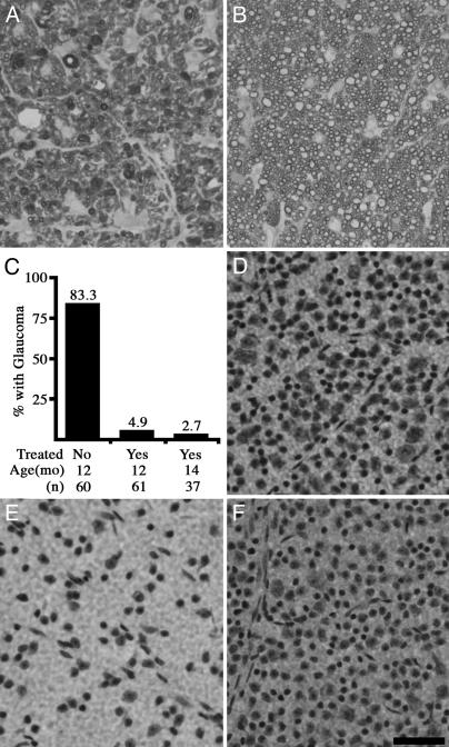
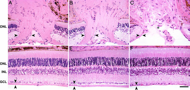
Here, we show that high-dose gamma-irradiation accompanied with syngeneic bone marrow transfer can confer complete protection against glaucoma in a mouse model. Because bone marrow genotype was unaltered by this procedure, it was not the causative agent. The neuroprotection is robust and highly reproducible. Glaucoma-prone DBA/2J mice received a single treatment at 5-8 weeks of age and were protected from glaucomatous retinal ganglion cell degeneration out to 14 months of age (oldest assessed). By 12-14 months, retinal ganglion cell degeneration is usually very severe and essentially complete in the majority of untreated DBA/2J mice. To assess reproducibility, three groups of mice were treated at different times, and the results were essentially the same each time. Considering all experiments, the vast majority of treated mice had no detectable glaucomatous neurodegeneration. A beneficial effect of treatment including high-dose radiation is unprecedented, and we are not aware of any other neuroprotective effects this substantial. Because of the robust protective effect, this treatment offers another tool for studying mechanisms of neuroprotection.


Figures

Fig. 1.
Fig. 1.
Clinical disease progression and glaucomatous insult is not influenced by radiation treatment. The overall clinical presentation of the iris disease is indistinguishable between treated and untreated groups. Typical images of mice of the indicated ages and treatment groups are shown. The only clinical difference in ocular phenotypes between untreated and treated cohorts was that all treated mice developed radiation induced lens opacities. (A and B) At 9 months, characteristic peripupillary swellings and dispersed pigment accumulations are evident in both treated and untreated mice. At this age, the degree of peripupillary iris atrophy (evident as white tissue adjacent to the pupil) varies from eye to eye in each treatment group. (C and D) At 12 months, dispersed pigment is clearly evident on the lens and across the surface of the iris. (E and F) At 14 months, there is advanced iris atrophy, which is not restricted to the peripupillary area. Full-thickness iris holes and severely atrophic areas that appear thin and depigmented occur in both groups. (G) IOP profiles showing that treatment did not change the glaucomatous IOP insult (mean ± SEM). The thickness of the gray line represents the mean IOP ± SEM (11.3 ± 0.25, n = 31) for DBA/2J mice at an age before ocular disease (3 months). The number of successful IOP recordings at each age are indicated.
Fig. 2.
Fig. 2.
Treated mice are protected from glaucomatous neurodegeneration. (A–C) Optic nerves are stained with paraphenylenediamine to visualize the myelin sheath of all axons, and differentially darkly stain the axoplasm of damaged and dying axons. This is an extremely sensitive technique that allows for the detection of a single sick/dying axon in the optic nerve. (A) By 12 months, the majority of optic nerves from untreated DBA/2J mice have severe glaucoma, as defined by massive axon loss. (B) The vast majority of optic nerves from treated mice had no detectable glaucomatous damage, even out to 14 months. (C) A summary of the data from 12- and 14-month-old mice clearly demonstrates the protective effect of treatment, which prevents glaucomatous neurodegeneration in the vast majority of eyes. Because the results did not differ, the data from the experiments at independent times are combined. (D–F) Nissl-stained flat-mounted retinas from position-matched regions of the superior peripheral retina also demonstrate the profound protective effect of treatment (n = 5 flat mounted retinas per group). (D) Young DBA/2J mouse showing normal density of ganglion cell layer cells before glaucomatous damage. (E) Twelve-month-old untreated DBA/2J mouse, showing substantial reduction in the number of soma as a result of glaucoma. (F) Twelve-month-treated DBA/2J mouse with normal number of soma. (Scale bar, 50 μm.)
Fig. 3.
Fig. 3.
Treatment prevents glaucomatous optic nerve excavation. (A) The optic nerve heads of control nonglaucomatous DBA/2J mice include large numbers of axons, as evidenced by a thick nerve fiber layer, entering the optic nerve head (nerve fiber layer on left side of optic nerve head is marked by arrowheads). (B) The thickness of the nerve fiber layer in treated DBA/2J mice (14-month-old example) is indistinguishable from nonglaucomatous controls. (C) In contrast, untreated DBA/2J mice have severe axon loss, as evidenced by a very atrophied nerve fiber layer. Their optic nerve heads are also severely excavated (asterisk), a hallmark of glaucoma (12-month example). (D–F) Position-matched images of retinal cross sections. (D) Nonglaucomatous DBA/2J control mouse. (E) Treated DBA/2J mouse, 14 months old. (F) Untreated DBA/2J mouse, 12 months old. The nerve fiber layer (arrowheads) is of normal thickness in treated DBA/2J retina (compare D with E) and severely atrophied in the untreated glaucomatous DBA/2J retina (compare D with F). There is an obvious loss of somas in the ganglion cell layer (GCL) of the untreated DBA/2J mouse (F) but not in the treated DBA/2J mouse (E; compare both to control retina in D). ONL, outer nuclear layer; INL, inner nuclear layer. (Scale bar, 50 μm.)

Similar articles

Cited by

References

    1. Ritch, R., Shields, M. B. & Krupin, T. (1996) The Glaucomas (Mosby, St. Louis).
    1. Weinreb, R. N. & Khaw, P. T. (2004) Lancet 363, 1711–1720. - PubMed
    1. Heijl, A., Leske, M. C., Bengtsson, B., Hyman, L. & Hussein, M. (2002) Arch. Ophthalmol. 120, 1268–1279. - PubMed
    1. Collaborative Normal-Tension Glaucoma Study Group (1998) Am. J. Ophthalmol. 126, 487–497. - PubMed
    1. Wax, M. B. & Tezel, G. (2002) Mol. Neurobiol. 26, 45–55. - PubMed

Publication types