Comparative Study
. 2005 Mar 9;25(10):2455-62.
doi: 10.1523/JNEUROSCI.4391-04.2005.

Certain inhibitors of synthetic amyloid beta-peptide (Abeta) fibrillogenesis block oligomerization of natural Abeta and thereby rescue long-term potentiation

Affiliations
Comparative Study

Certain inhibitors of synthetic amyloid beta-peptide (Abeta) fibrillogenesis block oligomerization of natural Abeta and thereby rescue long-term potentiation

Dominic M Walsh et al. J Neurosci. .

Erratum in

  • J Neurosci. 2005 May 4;25(18):4658

Abstract

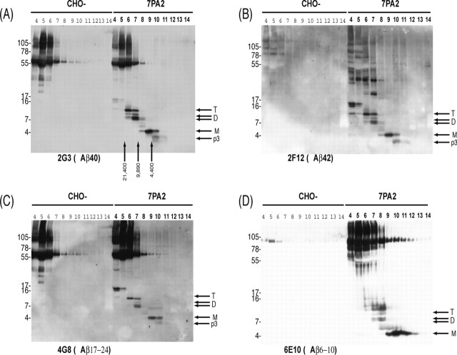
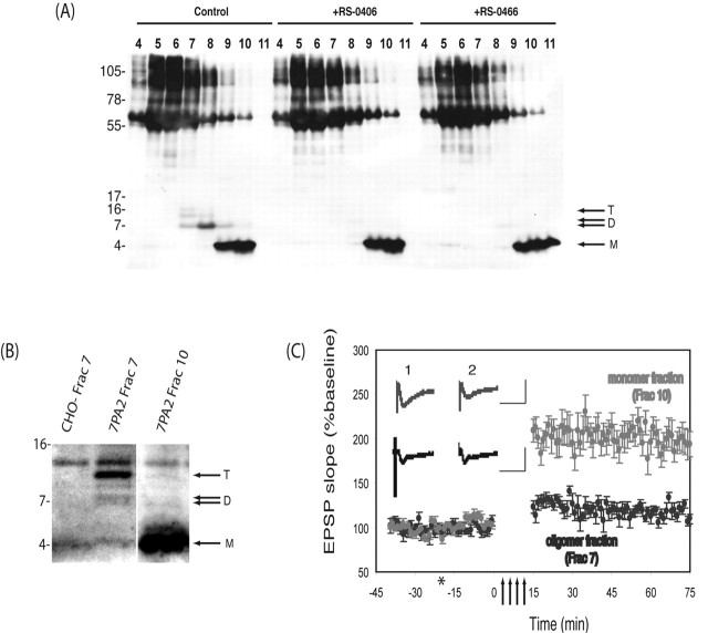
Recent studies support the hypothesis that soluble oligomers of amyloid beta-peptide (Abeta) rather than mature amyloid fibrils are the earliest effectors of synaptic compromise in Alzheimer's disease. We took advantage of an amyloid precursor protein-overexpressing cell line that secretes SDS-stable Abeta oligomers to search for inhibitors of the pathobiological effects of natural human Abeta oligomers. Here, we identify small molecules that inhibit formation of soluble Abeta oligomers and thus abrogate their block of long-term potentiation (LTP). Furthermore, we show that cell-derived Abeta oligomers can be separated from monomers by size exclusion chromatography under nondenaturing conditions and that the isolated, soluble oligomers, but not monomers, block LTP. The identification of small molecules that inhibit early Abeta oligomer formation and rescue LTP inhibition offers a rational approach for therapeutic intervention in Alzheimer's disease and highlights the utility of our cell-culture paradigm as a useful secondary screen for compounds designed to inhibit early steps in Abeta oligomerization under biologically relevant conditions.


Figures

Figure 1.
Figure 1.
Hydroxyanaline derivatives RS-0406 and RS-0466 inhibit Aβ oligomerization A, Synthetic Aβ(1-42) was incubated at 100 μm in 50 mm Tris-HCl, pH 7.4, at 37°C with and without 0.25 mm of each test compound, and aliquots were assayed for thioflavin T binding. RS-0406, OR1, and OR2 each significantly decreased in vitro synthetic fibrillogenesis (*p < 0.05). Error bars represent SD. B, 7PA2 cells were grown to near confluence in 10 cm2 dishes and allowed to condition serum-free medium with and without test compounds [(in μm): 102 RS-0406, 9.3 RS-0466, 18.5 OR1, and 18.5 OR2]. After 15 h, the CM was collected, cleared of cells, and analyzed by IP with R1282 and Western blotting with 6E10. Only RS-0406 and RS-0466 decreased natural oligomer levels. M, D, and T designate Aβ monomer, dimer, and trimer. C, 7PA2 cells were grown to near confluence in 10 cm2 dishes as described in B but labeled with [35S]methionine for 15 h with and without RS-0406 or RS-0466. Thereafter, the CM was collected, cleared of cells, immunoprecipitated with R1282, and analyzed by SDS-PAGE and autoradiography. M, D, and T are as in B. Aβ monomer, 5 kDa Aβ, and p3 (the latter being the proteolytic product of the α- and γ-cleavages of APP) do not appear as discrete bands at the exposure time necessary to visualize Aβ oligomers. Again RS-0466 and RS-0406 decreased oligomer levels. D, 7PA2 cells were grown in the presence or absence of RS-0406 or RS-0466, and lysates were prepared and immunoprecipitated with R1282. Both Aβ monomer (M) and dimer (D) bands are readily detected in lysates from untreated cells (0), but the dimer bands are significantly reduced in lysates of cells grown in the presence of RS-0406 or RS-0466. Because 6E10 also detects the more abundant APP C-terminal fragment, C99, the trimer is not discernible. Both compounds decreased the amount of dimers detected intracellularly. E, 7PA2 cells were grown to near confluence in 10 cm2 dishes as described in B and allowed to condition serum-free medium in the absence of any compounds. After harvesting and removing the cells by low-speed centrifugation, the CM was incubated with and without RS-0406 or RS-0466 for 15 h at 37°C. Neither compound altered the pattern of detection of oligomers, thus confirming that these hydroxyanaline compounds do not act by breaking up preexisting oligomers but rather act by inhibiting oligomerization.
Figure 2.
Figure 2.
Conditioned medium from 7PA2 cells treated with RS-0406 or RS-0466 do not alter hippocampal LTP. A, Perfusion of hippocampal slices with 7PA2 CM (blue), but not with CHO-CM (black), blocked LTP of excitatory synaptic transmission in the CA1 area [131.3 ± 8.3% at 60 min after HFS (indicated by arrows); mean ± SEM percentage of baseline; n = 6; p > 0.05 compared with pre-HFS baseline]. Insets show typical EPSPs ∼5 min pre- (1) and ∼60 min post-HFS (2). Calibration: 10 ms, 1 mV. Asterisk denotes addition of test media. B, Perfusion of hippocampal slices with CM from 7PA2 cells conditioned in the presence of RS-0406 (red) or RS-0466 (green) resulted in no inhibition of LTP (196.7 ± 11.3% at 60 min post-HFS, n = 7, p > 0.05; 196.7 ± 19.4% at 60 min post-HFS, n = 7, p > 0.05, respectively). Calibration: 25 ms, 2 mV. C, Perfusion of hippocampal slices with 7PA2 CM spiked acutely with RS-0406 (red open circles) or RS-0466 (green open circles) did not reverse the inhibition of LTP (113.9 ± 3.6% at 60 min post-HFS, n = 5, p > 0.05; 111.1 ± 8.4% at 60 min post-HFS, n = 7, p > 0.05, respectively), Calibration: 25 ms, 2 mV. D, Magnitude of LTP at 60 min post-HFS (percentage of baseline ± SEM). Treatments and the number of slices examined per treatment are indicated beneath each histogram. All treatments refer to the use of 7PA2 CM unless otherwise indicated. The term post indicates that compound was added to medium after it had been conditioned by cells, as opposed to addition before conditioning. Medium conditioned by 7PA2 cells in the presence of either RS-0406 or RS-0466 facilitated normal LTP, whereas addition of either compound after conditioning did not prevent the oligomer-mediated block of LTP. Error bars represent SEM.
Figure 3.
Figure 3.
Size exclusion fractionated oligomers are detected by antibodies to the mid region, N terminus, and C terminus of Aβ. Conditioned media from CHO and 7PA2 cells were concentrated ∼10-fold and then chromatographed on a Superdex 75 column, and 1 ml fractions were collected, lyophilized, and Western blotted. Lanes are labeled according to elution volume, and the elution of dextran standards is indicated by vertical arrows. Antibodies to the mid region (C; 4G8), N terminus (D; 6E10), and C terminus (A, 2G3; B, 21F12) of Aβ revealed the resolution of Aβ monomers (M), dimers (D), and trimers (T). The specificity of these bands is confirmed by the fact that they are only detected in fractions of 7PA2 CM (fraction numbers in bold type) and not in fractions of CHO-CM. Samples shown in A and B were from the same fractionation, and those shown in C and D were from separate fractionations. In each case, the blots shown are representative of at least three different experiments.
Figure 4.
Figure 4.
SEC-isolated Aβ oligomers, but not monomers, inhibit LTP. A, 7PA2 cells were allowed to condition serum-free medium in the presence or absence of RS-0406 or RS-0466 for 15 h, and the CM was concentrated as described. The concentrated CM was subjected to SEC, and fractions were analyzed by Western blotting using a combination of 2G3 and 21F12. Aβ monomers (M), dimers (D), and trimers (T) are indicated by arrows. B, SEC fractions containing monomeric or oligomeric Aβ were lyophilized and reconstituted in 10 ml of ACSF, and an aliquot (1.5 ml) was analyzed by IP/Western blot. C, Perfusion of hippocampal slices with oligomeric Aβ (fraction 7) inhibited LTP (115.8 ± 10.0% at 60 min post-HFS; mean ± SEM percentage of baseline; n = 6; p > 0.05 compared with pre-HFS baseline), whereas perfusion with monomeric Aβ (fraction 9) did not alter LTP (227.1 ± 22.6% at 60 min post-HFS; mean ± SEM percentage of baseline; n = 6; p > 0.05 compared with pre-HFS baseline). Insets show typical EPSPs ∼5 min pre- (1) and ∼60 min post-HFS (2). Calibration: 25 ms, 2 mV. Arrows signify the period of HFS, and the asterisk indicates addition of test media.

References

    1. Cohen FE, Kelly JW (2003) Therapeutic approaches to protein-misfolding diseases. Nature 426: 905-909. - PubMed
    1. Davies CA, Mann DM, Sumpter PQ, Yates PO (1987) A quantitative morphometric analysis of the neuronal and synaptic content of the frontal and temporal cortex in patients with Alzheimer's disease. J Neurol Sci 78: 151-164. - PubMed
    1. Findeis MA (2002) Peptide inhibitors of beta amyloid aggregation. Curr Top Med Chem 2: 417-423. - PubMed
    1. Haass C, Schlossmacher M, Hung AY, Vigo-Pelfrey C, Mellon A, Ostaszewski B, Lieberburg I, Koo EH, Schenk D, Teplow D, Selkoe D (1992) Amyloid β-peptide is produced by cultured cells during normal metabolism. Nature 359: 322-325. - PubMed
    1. Hardy J, Selkoe DJ (2002) The amyloid hypothesis of Alzheimer's disease: progress and problems on the road to therapeutics. Science 297: 353-356. - PubMed

Publication types

MeSH terms

LinkOut - more resources