A glial endogenous cannabinoid system is upregulated in the brains of macaques with simian immunodeficiency virus-induced encephalitis
- PMID: 15758162
- PMCID: PMC6725174
- DOI: 10.1523/JNEUROSCI.3923-04.2005
A glial endogenous cannabinoid system is upregulated in the brains of macaques with simian immunodeficiency virus-induced encephalitis
Erratum in
- J Neurosci. 2005 Mar 30;25(13):1 p following 3477. Kim, Wong-Ki [corrected to Kim, Woong-Ki];Williams, Ken [corrected to Williams, Kenneth]
Abstract
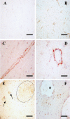
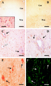
Recent evidence supports the notion that the endocannabinoid system may play a crucial role in neuroinflammation. We explored the changes that some elements of this system exhibit in a macaque model of encephalitis induced by simian immunodeficiency virus. Our results show that profound alterations in the distribution of specific components of the endocannabinoid system occur as a consequence of the viral infection of the brain. Specifically, expression of cannabinoid receptors of the CB2 subtype was induced in the brains of infected animals, mainly in perivascular macrophages, microglial nodules, and T-lymphocytes, most likely of the CD8 subtype. In addition, the endogenous cannabinoid-degrading enzyme fatty acid amide hydrolase was overexpressed in perivascular astrocytes as well as in astrocytic processes reaching cellular infiltrates. Finally, the pattern of CB1 receptor expression was not modified in the brains of infected animals compared with that in control animals. These results resemble previous data obtained in Alzheimer's disease human tissue samples and suggest that the endocannabinoid system may participate in the development of human immunodeficiency virus-induced encephalitis, because activation of CB2 receptors expressed by immune cells is likely to reduce their antiviral response and thus could favor the CNS entry of infected monocytes.
Figures


Similar articles
-
Expression of proinflammatory cytokines and its relationship with virus infection in the brain of macaques inoculated with macrophage-tropic simian immunodeficiency virus.Neuropathology. 2009 Feb;29(1):13-9. doi: 10.1111/j.1440-1789.2008.00929.x. Epub 2008 May 27. Neuropathology. 2009. PMID: 18507770
-
Cannabinoid CB2 receptors and fatty acid amide hydrolase are selectively overexpressed in neuritic plaque-associated glia in Alzheimer's disease brains.J Neurosci. 2003 Dec 3;23(35):11136-41. doi: 10.1523/JNEUROSCI.23-35-11136.2003. J Neurosci. 2003. PMID: 14657172 Free PMC article.
-
Cannabinoid receptor expression in HIV encephalitis and HIV-associated neuropathologic comorbidities.Neuropathol Appl Neurobiol. 2011 Aug;37(5):464-83. doi: 10.1111/j.1365-2990.2011.01177.x. Neuropathol Appl Neurobiol. 2011. PMID: 21450051 Free PMC article.
-
Searching for clues: tracking the pathogenesis of human immunodeficiency virus central nervous system disease by use of an accelerated, consistent simian immunodeficiency virus macaque model.J Infect Dis. 2002 Dec 1;186 Suppl 2:S199-208. doi: 10.1086/344938. J Infect Dis. 2002. PMID: 12424698 Review.
-
Role of the endocannabinoid system in Alzheimer's disease: new perspectives.Life Sci. 2004 Sep 3;75(16):1907-15. doi: 10.1016/j.lfs.2004.03.026. Life Sci. 2004. PMID: 15306158 Review.
Cited by
-
Inhibitory Neurotransmission Is Sex-Dependently Affected by Tat Expression in Transgenic Mice and Suppressed by the Fatty Acid Amide Hydrolase Enzyme Inhibitor PF3845 via Cannabinoid Type-1 Receptor Mechanisms.Cells. 2022 Mar 2;11(5):857. doi: 10.3390/cells11050857. Cells. 2022. PMID: 35269478 Free PMC article.
-
Functional selectivity in CB(2) cannabinoid receptor signaling and regulation: implications for the therapeutic potential of CB(2) ligands.Mol Pharmacol. 2012 Feb;81(2):250-63. doi: 10.1124/mol.111.074013. Epub 2011 Nov 7. Mol Pharmacol. 2012. PMID: 22064678 Free PMC article.
-
Neuroimmunity, drugs of abuse, and neuroAIDS.J Neuroimmune Pharmacol. 2006 Mar;1(1):41-9. doi: 10.1007/s11481-005-9001-3. J Neuroimmune Pharmacol. 2006. PMID: 18040790 Review.
-
Commentary: Functional Neuronal CB2 Cannabinoid Receptors in the CNS.Curr Neuropharmacol. 2011 Mar;9(1):205-8. doi: 10.2174/157015911795017416. Curr Neuropharmacol. 2011. PMID: 21886591 Free PMC article.
-
Cannabinoids inhibit migration of microglial-like cells to the HIV protein Tat.J Neuroimmune Pharmacol. 2011 Dec;6(4):566-77. doi: 10.1007/s11481-011-9291-6. Epub 2011 Jul 7. J Neuroimmune Pharmacol. 2011. PMID: 21735070
References
-
- Carrier EJ, Kearn CS, Barkmeier AJ, Breese NM, Yang W, Nithipatikom K, Pfister SL, Campbell WB, Hillard CJ (2004) Cultured rat microglial cells synthesize the endocannabinoid 2-arachidonylglycerol, which increases proliferation via a CB2 receptor-dependent mechanism. Mol Pharmacol 65: 999-1007. - PubMed
-
- Derocq JM, Jbilo O, Bouaboula M, Segui M, Clere C, Casellas P (2000) Genomic and functional changes induced by the activation of the peripheral cannabinoid receptor CB2 in the promyelocytic cells HL-60. Possible involvement of the CB2 receptor in cell differentiation. J Biol Chem 275: 15621-15628. - PubMed
-
- Felder CC, Nielsen A, Briley EM, Palkovits M, Priller J, Axelrod J, Nguyen DN, Richardson JM, Riggin RM, Koppel GA, Paul SM, Becker GW (1996) Isolation and measurement of the endogenous cannabinoid receptor agonist, anandamide, in brain and peripheral tissues of human and rat. FEBS Lett 393: 231-235. - PubMed
Publication types
MeSH terms
Substances
Grants and funding
LinkOut - more resources
Full Text Sources
Other Literature Sources
Research Materials
