Comparative Study
. 2005 Mar 9;25(10):2566-75.
doi: 10.1523/JNEUROSCI.4998-04.2005.

Beta-amyloid-stimulated microglia induce neuron death via synergistic stimulation of tumor necrosis factor alpha and NMDA receptors

Affiliations
Comparative Study

Beta-amyloid-stimulated microglia induce neuron death via synergistic stimulation of tumor necrosis factor alpha and NMDA receptors

Angela M Floden et al. J Neurosci. .

Abstract

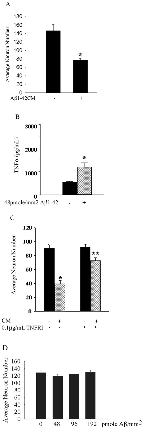
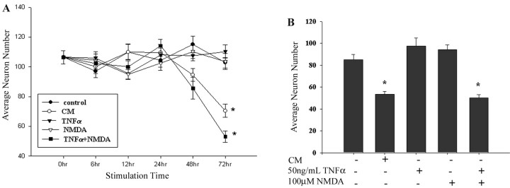
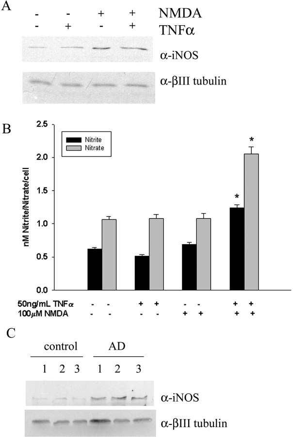
Although abundant reactive microglia are found associated with beta-amyloid (Abeta) plaques in Alzheimer's disease (AD) brains, their contribution to cell loss remains speculative. A variety of studies have documented the ability of Abeta fibrils to directly stimulate microglia in vitro to assume a neurotoxic phenotype characterized by secretion of a plethora of proinflammatory molecules. Collectively, these data suggest that activated microglia play a direct role in contributing to neuron death in AD rather than simply a role in clearance after plaque deposition. Although it is clear the Abeta-stimulated microglia acutely secrete toxic oxidizing species, the identity of longer-lived neurotoxic agents remains less defined. We used Abeta-stimulated conditioned media from primary mouse microglia to identify more stable neurotoxic secretions. The NMDA receptor antagonists memantine and 2-amino-5-phosphopetanoic acid as well as soluble tumor necrosis factor alpha (TNFalpha) receptor protect neurons from microglial-conditioned media-dependent death, implicating the excitatory neurotransmitter glutamate and the proinflammatory cytokine TNFalpha as effectors of microglial-stimulated death. Neuron death occurs in an oxidative damage-dependent manner, requiring activity of inducible nitric oxide synthase. Toxicity results from coincident stimulation of the TNFalpha and NMDA receptors, because stimulations of either alone are insufficient to initiate cell death. These findings suggest the hypothesis that AD brains provide the appropriate microglial-mediated inflammatory environment for TNFalpha and glutamate to synergistically stimulate toxic activation of their respective signaling pathways in neurons as a contributing mechanism of cell death.


Figures

Figure 1.
Figure 1.
Conditioned media from Aβ-stimulated microglia induce neuron death in a TNFα-dependent manner. Conditioned Neurobasal media (CM) from primary mouse microglia (400 cells/mm2) stimulated for 48 h with or without (control) immobilized Aβ1-42 fibrils (48 pmol/mm2) was generated. A, Control and Aβ-stimulated conditioned media were applied to mouse cortical neuron cultures (E16; 7 d in vitro) for 72 h. Neurons were fixed in 4% paraformaldehyde, stained using anti-MAP2 antibody, and counted. Neurons from four fields/conditions were counted in quadruplicate wells and averaged ± SEM. The graph is representative of three independent experiments. B, Conditioned media were collected, and TNFα concentrations were determined via commercial ELISA. The graph is representative of three independent experiments. C, Cortical neurons were cultured for 72 h with unstimulated and Aβ-stimulated conditioned medium in the absence or presence of soluble TNFRI (0.1 μg/ml). Neurons were fixed, stained using anti-MAP2 antibody, counted as above, and averaged ± SEM. The graph is representative of three independent experiments. D, Media from plates containing increasing concentrations of immobilized Aβ (0, 48, 96, and 192 pmol Aβ1-42/mm2) alone were transferred to cortical neurons for 72 h. Neurons were fixed and counted, and statistical significance was determined as above. *p < 0.001 from control; **p < 0.001 from CM.
Figure 2.
Figure 2.
Conditioned media-dependent death requires NMDA receptor activity. A, Conditioned Neurobasal media (CM) from primary mouse microglia (400 cells/mm2) stimulated for 48 h with or without (control) immobilized Aβ1-42 fibrils (48 pmol/mm2) were generated, and micromolar glutamate concentrations were calculated from the media. The graph is the average ± SEM of three independent experiments. Cortical neurons (E16; 7 d in vitro) were cultured for 72 h with conditioned medium from primary mouse microglia that were unstimulated or stimulated (CM) 48 h with Aβ1-42 fibrils in the absence or presence of 1 μm memantine and 10 μm APV (B) or 1, 5, and 50 μm NBQX (C). Neurons were then fixed, stained using anti-MAP2 antibody, and counted. Neurons from four fields/conditions were counted in quadruplicate wells and averaged ± SEM. Graphs are representative of three independent experiments. *p < 0.001 from control; **p < 0.001 from CM.
Figure 3.
Figure 3.
TNFα and glutamate/NMDA synergistically stimulate neuron death. Mouse cortical neurons (E16; 7 d in vitro) were cultured in the absence or presence of NMDA (50, 100 μm) (A), glutamate (25, 50, 100 μm) (B), and mouse TNFα (5, 50 ng/ml). Stimuli were added to neurons for 72 h and then cells were fixed, stained using anti-MAP2 antibody, and counted. Neurons from four fields/conditions were counted in quadruplicate wells and averaged ± SEM. Graphs are representative of three independent experiments. *p < 0.05 from control; **p < 0.001 from control.
Figure 4.
Figure 4.
Conditioned media and TNFα plus NMDA-dependent neuron death have a similar time course. Mouse cortical neurons (E16; 7 d in vitro) were cultured in the absence or presence of conditioned medium from microglia that were unstimulated or stimulated (CM) 48 h with Aβ1-42 fibrils, NMDA (100 μm), and mouse TNFα (50 ng/ml). A, Stimuli were added to neurons for 0-72 h. At select time points, cells were fixed, stained, and counted. B, Alternatively, stimuli were added to neurons for 1 h and then replaced with fresh media and fixed after a total time of 72 h. Neurons from four fields/conditions were counted in quadruplicate wells and averaged ± SEM. Graphs are representative of three independent experiments. *p < 0.001 from control.
Figure 5.
Figure 5.
Conditioned media and TNFα plus NMDA-dependent neuron death both involve iNOS activity and peroxynitrite formation. Mouse cortical neurons (E16; 7 d in vitro) were cultured in the absence or presence of conditioned medium from primary mouse microglia that were unstimulated or stimulated (CM) 48 h with Aβ1-42 fibrils, NMDA (100 μm), and mouse TNFα (50 ng/ml). A, Conditioned media were added to neurons in the absence or presence of 100 μm MnTBAP or 10 μm 1400W.2HCl. B, TNFα and/or NMDA was added to neurons in the absence or presence of 100 μm MnTBAP or 10 μm 1400W.2HCl. Neurons were stimulated for 72 h and then fixed, stained, and counted. Neurons from four fields/conditions were counted in quadruplicate wells and averaged ± SEM. Graphs are representative of three independent experiments. *p < 0.001 from control; **p < 0.001 from CM.
Figure 6.
Figure 6.
TNFα plus NMDA-dependent neuron death stimulates increased iNOS protein levels and activity. Neurons were unstimulated or stimulated 72 h with TNFα (50 ng/ml), 100 μm NMDA, or TNFα plus NMDA. A, To determine iNOS protein levels, neurons were lysed, and proteins were resolved by 10% SDS-PAGE and Western blotted using rabbit anti-iNOS antibody. Antibody binding was visualized by enhanced chemiluminescence. Lysates were blotted with antibody recognizing neuron-specific βIII tubulin as a protein-loading control. Blots are representative of four independent experiments. B, To assess nitric oxide synthase, activity concentrations of nitrite and nitrate in neuronal media were quantitated after 72 h of stimulation with TNFα, NMDA, or TNFα plus NMDA. Nitrite/nitrate concentrations were normalized to cell number and averaged from six repeats per condition from three independent experiments (*p < 0.001 compared with respective control). C, Proteins from three AD and three control brain midtemporal gyrus homogenates were separated by 10% SDS-PAGE and Western blotted using rabbit anti-iNOS antibody. Antibody binding was visualized by enhanced chemiluminescence. Lysates were blotted with antibody recognizing neuron-specific βIII tubulin as a protein-loading control.
Figure 7.
Figure 7.
TNFα plus NMDA-dependent neuron death stimulates increased protein nitrotyrosine levels. Neurons were unstimulated or stimulated 72 h with TNFα (50 ng/ml), 100 μm NMDA, TNFα plus NMDA, or 1 and 5 μm SIN-1. A, To determine nitrotyrosine protein levels, neurons were lysed, and proteins were immunoprecipitated and resolved by 10% SDS-PAGE and Western blotted using mouse anti-nitrotyrosine antibodies. Antibody binding was visualized by enhanced chemiluminescence. Blots are representative of four independent experiments. B, To assess SIN-1-dependent protein, nitration cells were lysed, and nitrated proteins were immunoprecipitated, resolved by 10% SDS-PAGE, and blotted with anti-nitrotyrosine antibody. C, Nitrotyrosine-containing proteins were immunoprecipitated from midtemporal gyrus homogenate from three AD and three control brain samples and separated by 10% SDS-PAGE and Western blotted using anti-nitrotyrosine antibody. Antibody binding was visualized by enhanced chemiluminescence. IgG heavy chain is identified (arrows) on the blots simply for orientation purposes.
Figure 8.
Figure 8.
TNFα plus NMDA induces increased neuronal iNOS immunoreactivity. Neurons were unstimulated (A-D) or stimulated (E-H) 72 h with TNFα (50 ng/ml) plus 100 μm NMDA and then fixed in 4% paraformaldehyde. Cultures were double labeled using anti-MAP2 and anti-iNOS antibodies with FITC and Texas Red-conjugated secondary antibodies, respectively. Cultures were mounted in DAPI containing mounting media for confocal imaging. A, E, Anti-MAP2; B, F, anti-iNOS; C, G, DAPI; D, merge of A-C; H, merge of E-G. Images are representative of three independent experiments.
Figure 9.
Figure 9.
Neuronal NMDAR subunit immunoreactivity colocalizes with TNFRI and TNFRII. Cortical neuron cultures from E16 mice were cultured for 7 d in vitro and then collected in RIPA buffer. A, Lysates were resolved by 10% SDS-PAGE and Western blotted using antibodies recognizing NMDAR1, TNFRI, and TNFRII. Antibody binding was visualized via enhanced chemiluminescence. B, Neurons were fixed at 7 d in vitro and dually immunostained using anti-TNFRI/TNFRII and anti-NMDAR1 antibodies. Primary antibody binding was visualized using Texas Red-conjugated anti-rabbit and FITC-conjugated anti-goat antibodies for TNFRI and TNFRII, respectively, and Vector VIP for NMDAR1. Arrows demonstrate examples of varying levels of immunoreactivity for each antigen.

Similar articles

Cited by

References

    1. Akiyama H, Barger S, Barnum S, Bradt B, Bauer J, Cole GM, Cooper NR, Eikelenboom P, Emmerling M, Fiebich BL, Finch CE, Frautschy S, Griffin WS, Hampel H, Hull M, Landreth G, Lue L, Mrak R, Mackenzie IR, McGeer PL, et al. (2000) Inflammation and Alzheimer's disease. Neurobiol Aging 21: 383-421. - PMC - PubMed
    1. Barger S, Basile AS (2001) Activation of microglia by secreted amyloid precursor protein evokes release of glutamate by cystine exchange and attenuates synaptic function. J Neurochem 76: 846-854. - PubMed
    1. Barger SW, Hortser D, Furukawa K, Goodman Y, Krieglstein J, Mattson MP (1995) Tumor necrosis factors alpha and beta protect neurons against amyloid beta-peptide toxicity: evidence for involvement of a kappa B-binding factor and attenuation of peroxide and Ca2+ accumulation. Proc Natl Acad Sci USA 92: 9328-9332. - PMC - PubMed
    1. Beckman J, Ischiropoulos H, Zhu L, van der Woerd M, Smith C, Chen J, Harrison J, Martin J, Tsai M (1992) Kinetics of superoxide dismutase- and iron-catalyzed nitration of phenolics by peroxynitrite. Arch Biochem Biophys 298: 438-445. - PubMed
    1. Bianca VD, Dusi S, Bianchini E, Dal Pra I, Rossi F (1999) Beta-amyloid activates the O-2 forming NADPH oxidase in microglia, monocytes, and neutrophils. A possible inflammatory mechanism of neuronal damage in Alzheimer's disease. J Biol Chem 274: 15493-15499. - PubMed

Publication types

MeSH terms

LinkOut - more resources