Comparative Study
. 2005 Mar 9;25(10):2576-85.
doi: 10.1523/JNEUROSCI.4118-04.2005.

Multiple interacting sites of ectopic spike electrogenesis in primary sensory neurons

Affiliations
Comparative Study

Multiple interacting sites of ectopic spike electrogenesis in primary sensory neurons

Ron Amir et al. J Neurosci. .

Abstract

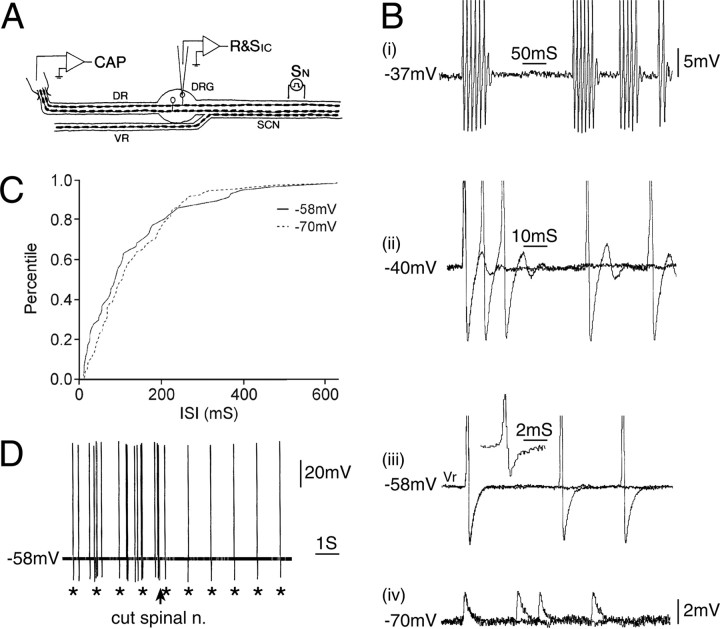
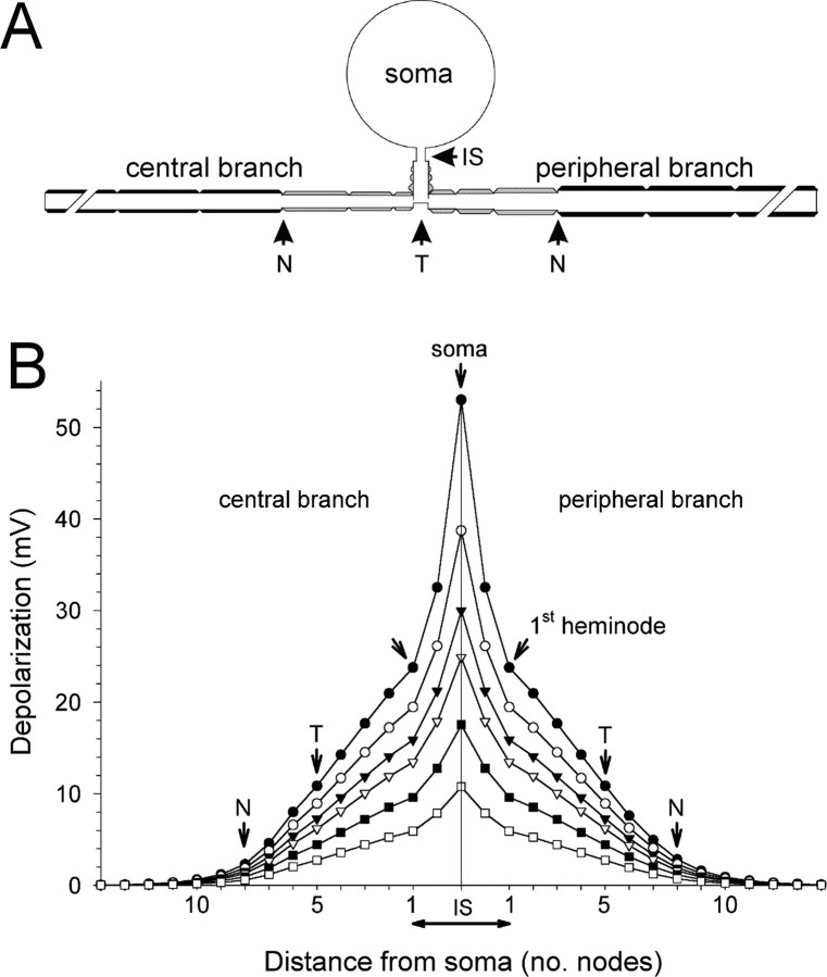
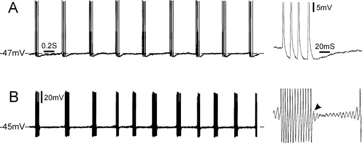
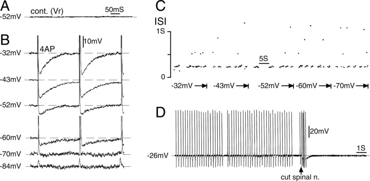
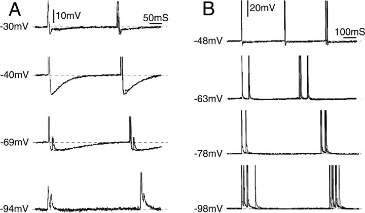
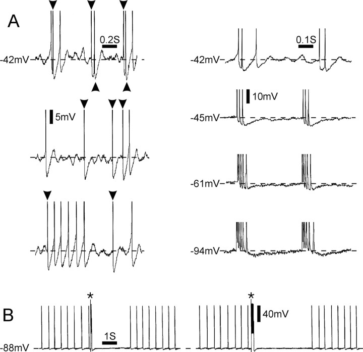
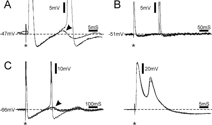
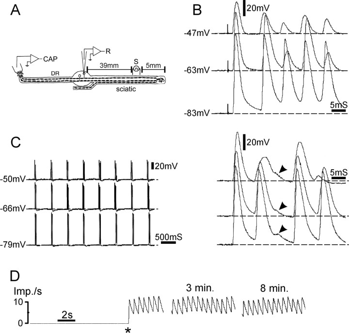
Ectopic discharge generated in injured afferent axons and cell somata in vivo contributes significantly to chronic neuropathic dysesthesia and pain after nerve trauma. Progress has been made toward understanding the processes responsible for this discharge using a preparation consisting of whole excised dorsal root ganglia (DRGs) with the cut nerve attached. In the in vitro preparation, however, spike activity originates in the DRG cell soma but rarely in the axon. We have now overcome this impediment to understanding the overall electrogenic processes in soma and axon, including the resulting discharge patterns, by modifying the bath medium in which recordings are made. At both sites, bursts can be triggered by subthreshold oscillations, a phasic stimulus, or spikes arising elsewhere in the neuron. In the soma, once triggered, bursts are maintained by depolarizing afterpotentials, whereas in the axon, an additional process also plays a role, delayed depolarizing potentials. This alternative process appears to be involved in "clock-like" bursting, a discharge pattern much more common in axons than somata. Ectopic spikes arise alternatively in the soma, the injured axon end (neuroma), and the region of the axonal T-junction. Discharge sequences, and even individual multiplet bursts, may be a mosaic of action potentials that originate at these alternative electrogenic sites within the neuron. Correspondingly, discharge generated at these alternative sites may interact, explaining the sometimes-complex firing patterns observed in vivo.


Figures

Figure 2.
Figure 2.
Spontaneous discharge originating in the axon rather than in the cell soma. A, The experimental setup. CAP, Suction electrode recording compound action potential; R&SIC, intracellular recording micropipette; SCN, sciatic nerve; SN, stimulating electrode on nerve. B, This neuron fired spontaneous spikes at Vr (-58 mV) but did not have subthreshold oscillations. Depolarization induced the appearance of a DAP after each spike, which sometimes triggered subsequent spikes, resulting in burst firing (traces at -37 and -40 mV). Hyperpolarization to -70 mV caused failure of soma spikes, leaving only residual (axonal) m-spikes. Thus, the soma spikes (singlets and bursts) at more depolarized potentials were triggered by action potentials of axonal origin. These axonal spikes also propagated into the L5 DR as demonstrated by the spike-triggered average of the signal recorded on the DR suction electrode (inset adjacent to the trace at -58 mV). C, Consistent with this interpretation, the frequency of the singlet spikes at -58 mV matched that of m-spikes at -70 mV (and spike bursts at -37 mV). The plot shows the cumulative distribution of singlet spike and m-spike ISI (d = 0.16; p > 0.2; K-S test; http://www.physics.csbsju.edu/stats/KS-test.html). D, Electrical stimulus pulses delivered to the spinal nerve (1 Hz; asterisks) triggered soma spikes. These summed with the spontaneously occurring spikes. Transection of the spinal nerve just distal to the stimulating electrode (arrow) eliminated the spontaneous spikes but not the stimulation-evoked spikes.
Figure 5.
Figure 5.
Simulation of the membrane potential decay from the soma to the T-stem and the central and peripheral branches. A, A sketch of the cell model. B, Depolarizing pulses (200 ms) were applied to the soma (current clamp), and the level of resultant depolarization at steady state was recorded in subsequent nodes along the T-stem and the central and peripheral axonal branches. IS, Initial segment, the unmyelinated part of the T-stem; T, T-junction. The three internodes between T and N in both peripheral and central branches were short and had fewer myelin wraps. Subsequent internodes had “normal” parameters (Amir and Devor, 2003a).
Figure 1.
Figure 1.
Clock-like versus irregular bursting. A, This cell fired multiplet bursts consisting of four spikes per burst, with clock-like regularity in the presence of 4-AP. A single burst is shown on an expanded time scale and gain on the right. Bursts were not preceded by subthreshold oscillations and were not followed by a DAP. Note the regular interval between multiplets (variability index, 0.02). B, Burst firing in a different cell was triggered by subthreshold oscillations and maintained by DAPs (B, right arrowhead). The interval between bursts was not as regular as the cell in A (variability index, 0.3). Both cells had a highly regular ISI within bursts (variability index: A, 0.08; B, 0.06). Calibration bars refer to both A and B.
Figure 3.
Figure 3.
Repetitive firing after 4-AP application originated in the nerve-end neuroma. A, This neuron, which was silent at Vr(-52 mV), began to fire bursts (with occasional misses) after 4-AP application (1 mm). cont., Control. B, Shifting the membrane potential between -32 and -84 mV had no effect on the ISI within a burst, although the AHP became smaller with hyperpolarization, and eventually the soma spike blocked, leaving residual m-spikes (at -84 mV). C, The voltage independence of firing frequency is also illustrated in this ISI dot raster plot. Dots above the main scatter at ISI = 215 ms (corresponding to a firing frequency of 4.7 Hz) are integer multiples of the basic ISI, indicating that they represent spikes missed within the burst (Matzner and Devor, 1993). D, Transection of the spinal nerve (arrow) eliminated the spiking, affirming its origin in the distal part of the axon.
Figure 4.
Figure 4.
Evidence for ectopic electrogenesis in the neuronal stem axon or T-junction. A, Tonic discharge emerged in this neuron after application of 4-AP. Subthreshold oscillations are absent; the spikes were triggered from outside of the cell soma (i.e., from the axon). However, the interval between adjacent spikes in the train (ISI) varied with the somatic membrane potential (range, -30 to -94 mV), indicating that the axonal site of spike initiation was within a few space-constants of the soma (hence the stem axon or T-junction). When the cell was hyperpolarized (-94 mV), soma spikes failed, leaving residual m-spikes. Note that at the axonal site of electrogenesis, firing was frequently in doublets (interval, 15 ms) but only at more hyperpolarized potentials. The first spike of each doublet triggered a soma spike; the second appeared as an m-spike within the AHP of the soma spike. B, In a second neuron that also fired tonically after 4-AP application, shifting Vm in the hyperpolarizing direction (from -48 to -98 mV) caused a shift from singlet to multiplet discharge and a progressive increase in the interval between bursts. ISI within multiplets, in contrast, decreased with hyperpolarization, and the number of spikes per multiplet increased. All traces show three superimposed sweeps.
Figure 6.
Figure 6.
Interaction among alternative sources of repetitive firing. A, Left, Spontaneous activity of singlets and short bursts recorded from the soma of a DRG neuron ∼6 min after 4-AP application. This cell exhibited both oscillations and DAPs only after 4-AP superfusion. Some of the spikes were generated in the soma by oscillations and DAPs (unmarked spikes), whereas others (arrowheads) invaded the soma after generation elsewhere. Spikes are truncated. Right, Hyperpolarization abolished oscillations, DAPs, and resultant soma spikes, leaving only invading spikes. These were generated in a tonic pattern with multiplets. After additional hyperpolarization, the ISI within a multiplet was gradually decreased, and the number of impulses increased; however, intermultiplet interval was unaffected.B, Tonic spontaneous activity in the absence of oscillations and DAPs, indicating that the firing was generated outside the soma. A 100 ms (left) or 200 ms (right) suprathreshold depolarizing pulse applied via the recording micropipette (asterisks) generated a burst of soma spikes and then abolished firing for several seconds, too long to be accounted for by spike collision. The spike burst apparently silenced electrogenesis at the axonal pacemaker site.
Figure 7.
Figure 7.
Afterdischarge triggered by stimulus pulses applied to the nerve (A, C) or the soma (B; asterisks) in the presence of 4-AP. A-C show three different neurons. A, Afterdischarge maintained by a postspike DAP (arrowhead). B, Afterdischarge triggered in the absence of a DAP, long after recovery from a prolonged AHP (3 traces; in 1 trace, afterdischarge was not evoked). C, In this neuron, axonal stimulation evoked two bursts of doublets. The first spike in the second doublet was triggered by a DAP (C, left, arrowhead). The second spike, however, appears during the falling phase of the first spike as seen on a faster time scale (C, right). In all panels but C (right), spikes are truncated. Two sweeps are superimposed in A, and three sweeps are superimposed in B and C.
Figure 8.
Figure 8.
Afterdischarge of prolonged rhythmic multiplets after chronic nerve lesion in the presence of 4-AP. A, The experimental setup. CAP, Suction electrode recording compound action potential. B, C, In this cell, a single stimulus pulse triggered a prolonged train of rhythmic multiplets, each multiplet consisting of up to six spikes. Traces in B show the first multiplet after the sciatic nerve stimulus at a variety of membrane potentials. C (right) shows subsequent multiplets. Changing membrane potential did not affect ISI between or within multiplets, indicating that they were generated at the cut nerve end. Note that the third spike was aborted (arrowheads). D, Firing rate plot shows that this afterdischarge burst pattern persisted for many minutes after the initiating stimulus pulse (asterisk). Imp./s, Impulses per second.

References

    1. Amir R, Devor M (1996) Chemically-mediated cross-excitation in rat dorsal root ganglia. J Neurosci 16: 4733-4741. - PMC - PubMed
    1. Amir R, Devor M (1997) Spike-evoked suppression and burst patterning in dorsal root ganglion neurons. J Physiol (Lond) 501: 183-196. - PMC - PubMed
    1. Amir R, Devor M (2003a) Electrical excitability of the soma of sensory neurons is required for spike invasion of the soma, but not for through-conduction. Biophys J 84: 2181-2191. - PMC - PubMed
    1. Amir R, Devor M (2003b) Extra spike formation in sensory neurons and the disruption of afferent spike patterning. Biophys J 84: 2700-2708. - PMC - PubMed
    1. Amir R, Michaelis M, Devor M (1999) Membrane potential oscillations in dorsal root ganglion neurons: role in normal electrogenesis and in neuropathic pain. J Neurosci 19: 8589-8596. - PMC - PubMed

Publication types

LinkOut - more resources