Review
. 2005 Apr;3(4):349-58.
doi: 10.1038/nrmicro1131.

Opinion: Cell entry machines: a common theme in nature?

Affiliations
Review

Opinion: Cell entry machines: a common theme in nature?

Michèle A Barocchi et al. Nat Rev Microbiol. 2005 Apr.

Abstract

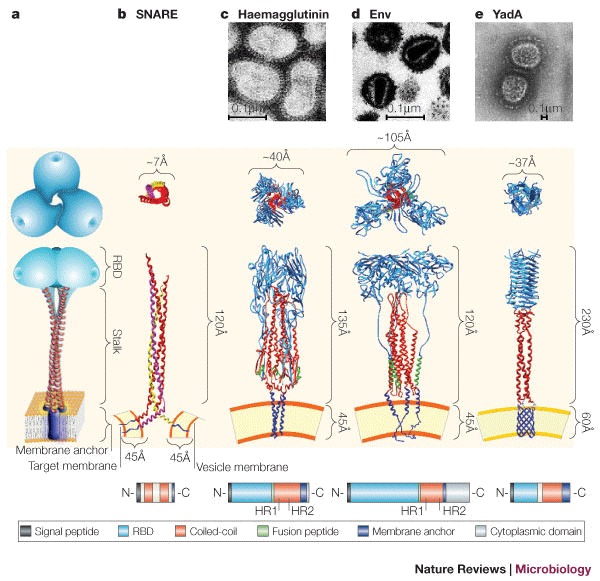
Molecular machines orchestrate the translocation and entry of pathogens through host cell membranes, in addition to the uptake and release of molecules during endocytosis and exocytosis. Viral cell entry requires a family of glycoproteins, and the structural organization and function of these viral glycoproteins are similar to the SNARE proteins, which are known to be involved in intracellular vesicle fusion, endocytosis and exocytosis. Here, we propose that a family of bacterial membrane proteins that are responsible for cell-mediated adherence and entry resembles the structural architecture of both viral fusion proteins and eukaryotic SNAREs and might therefore share similar, but distinct, mechanisms of cell membrane translocation. Furthermore, we propose that the recurrence of these molecular machines across species indicates that these architectural motifs were evolutionarily selected because they provided the best solution to ensure the survival of pathogens within a particular environment.


Conflict of interest statement

The authors are employed by Chiron Vaccines

Figures

Figure 1
Figure 1. Schematic representation of intracellular vesicle exocytosis, and viral and bacterial cell entry.
a | Attachment of intracellular vesicles to the plasma membrane is mediated by vesicle and target SNAREs (vSNAREs and tSNAREs, respectively), viral fusion proteins and bacterial invasins. b | Assembly and conformational changes of surface proteins result in the formation of a coiled-coil metastable structure, which induces tethering, followed by intimate adherence of vesicles and pathogens to the plasma membrane. In the case of HIV, the fusion protein undergoes a conformational change that results in insertion of the fusion peptide (shown in black) into the plasma membrane. Influenza virus entry is pH dependent — the virus is internalized and the acidification of the endosome results in a conformational change of haemagglutinin. This exposes the fusion peptide, which then inserts into the vesicular membrane, causing fusion of the viral and endosomal membrane. c | Energy derived from the conformational changes in b promote either fusion of cellular membranes (as seen for both vesicles and viruses) or invasion of host cells by bacteria. SNARE, soluble NSF (N-ethylmaleimide-sensitive fusion protein) accessory protein (SNAP) receptor.
Figure 2
Figure 2. Architecture of eukaryotic membrane and viral fusion proteins compared with bacterial invasins.
a | Schematic representation of the common trimeric structure consisting of a membrane anchor (purple), a central coiled-coil motif (stalk, red) and a globular receptor-binding domain (RBD, cyan). b | Partial three-dimensional structure of the coiled-coil domains of the synaptic SNARE complex. Synaptobrevin 2 (also known as VAMP2) (yellow) is anchored to the vesicle membrane. Syntaxin (pink) and SNAP25 (red) are anchored to the target membrane. These proteins are anchored to the cytoplasmic membrane by a hydrophobic α-helical C- terminal domain, which allows the protein to extend 120Å from the cell membrane. c | Structural representation of the influenza haemagglutinin (HA) trimer with the fusion peptide (green). The stalk is in its metastable pre-fusogenic state. The globular receptor-binding subunit HA1 is shown in blue, and the triple-stranded, coiled-coil fibrous region, known as HA2, is shown in red. d | The HIV envelope protein (Env) is composed of two non-covalently associated trimeric gp120 (red) and gp41 (blue) subunits. Here, gp120 is artificially fused to gp41. The fusion peptide, which is located in gp41, is depicted in green. In their monomeric form, both fusion proteins — haemagglutinin and Env — span the viral envelope with a single transmembrane α-helix near the C-terminus. e | Structural model of the trimeric Yersinia spp. YadA molecule anchored to the bacterial outer membrane through a β-barrel (blue). The anchor domain is represented by a porin-like outer membrane protein. Top–down views of the heterotrimeric SNARE complex, homotrimeric viral and bacterial fusion proteins (invasins), are shown. Structures are not to scale. The structures used to build the models have been obtained from the PDB protein databank (see the Online links box). Short domains for which coordinates were not available were built manually on the basis of secondary structure predictions. The anchor domain was modelled on the integral outer membrane protein (OmpA) from Escherichia coli (strain 1BXW), whereas the coiled-coil stalk was modelled on the structure of chicken cartilage trix protein (1AQ5). Transmission electron microscopy (TEM) of negatively stained influenza virus (c), HIV (d) and Yersinia spp. (e) are shown. The bottom panel is a schematic representation of the protein sequence architecture of the corresponding proteins. HR1 and HR2 are experimentally determined heptad repeats for haemagglutinin and Env. White boxes represent sequence portions with an undefined role. SNARE, soluble NSF (N-ethylmaleimide-sensitive fusion protein) accessory protein (SNAP) receptor. Panel c TEM courtesy of Linda M. Stannard, University of Cape Town, South Africa. Panel c (structural image) reproduced, with permission, from Ref. © (2002) Elsevier Science. Panel d TEM courtesy of H. Gelderblom, Koch Institute, Berlin. Panel e TEM reproduced, with permission, from Ref. © (2000) European Molecular Biology Organization.
Figure 3
Figure 3. Intracellular signalling events.
The signalling cascades and intracellular pathways that are used by viruses and bacteria to induce host cell uptake converge on those used in vesicle trafficking. A common theme is the activation of GTPases and the subsequent cytoskeletal changes that aid in the movement of vesicles or in the uptake of pathogens. a | In SNARE-mediated vesicle trafficking and exocytosis, intracellular factors such as the exocyst protein complex and the small GTPase Rab3A are important in the docking of the vesicle to the plasma membrane. Vesicle trafficking also involves the activation of intracellular cytoskeleton effector molecules such as phosphatidylinositol 3-kinase (PI3K) and other small GTPases such as Cdc42. F-actin nucleation mediated by N-WASP (neural Wiskott–Aldrich syndrome protein) and ARP2/3, and microtubule elongation are crucial in trafficking events. Receptor-mediated attachment of viruses such as influenza virus (b) and bacteria such as Yersinia spp. (c) also leads to the signalling and activation of GTPases (such as dynamin, Ras, Rac1 and Cdc42), as well as actin polymerization through the ARP2/3 complex (mediated by N-WASP in bacteria, and N-WASPand intersectin in viruses). PI3K is activated by the binding of certain viruses and bacteria to cell surface receptors, and is also implicated in actin polymerization. AP2, adaptor protein-2; CAS, Crk-associated substrate; CAT2, cationic amino-acid transporter; DAG, diacylglycerol; FAK, focal adhesion kinase; GRB2, growth factor receptor-bound protein-2; MAPK, mitogen-activated protein kinase; PIP2, phosphatidylinositol-4,5-bisphosphate; PI4P, phosphatidylinositol 4-phosphate; PKC, protein kinase C; SH2, Src-homology domain-2; SNARE, soluble NSF (N-ethylmaleimide-sensitive fusion protein) accessory protein (SNAP) receptor.

References

    1. Sutton RB, Fasshauer D, Jahn R, Brunger AT. Crystal structure of a SNARE complex involved in synaptic exocytosis at 2. 4Å resolution. Nature. 1998;395:347–353. doi: 10.1038/26412. - DOI - PubMed
    1. Antonin W, Fasshauer D, Becker S, Jahn R, Schneider TR. Crystal structure of the endosomal SNARE complex reveals common structural principles of all SNAREs. Nature Struct. Biol. 2002;9:107–111. doi: 10.1038/nsb746. - DOI - PubMed
    1. Chen YA, Scheller RH. Snare-mediated membrane fusion. Nature Rev. Mol. Cell Biol. 2001;2:98–106. doi: 10.1038/35052017. - DOI - PubMed
    1. Weber T, et al. SNAREpins: minimal machinery for membrane fusion. Cell. 1998;92:759–772. doi: 10.1016/S0092-8674(00)81404-X. - DOI - PubMed
    1. Dormitzer PR, Nason EB, Prasad BV, Harrison SC. Structural rearrangements in the membrane penetration protein of a non-enveloped virus. Nature. 2004;430:1053–1058. doi: 10.1038/nature02836. - DOI - PMC - PubMed

MeSH terms