

Assessment of the paraspinal muscles of subjects presenting an idiopathic scoliosis: an EMG pilot study
- PMID: 15760468
- PMCID: PMC1079862
- DOI: 10.1186/1471-2474-6-14
Assessment of the paraspinal muscles of subjects presenting an idiopathic scoliosis: an EMG pilot study
Abstract
Background: It is known that the back muscles of scoliotic subjects present abnormalities in their fiber type composition. Some researchers have hypothesized that abnormal fiber composition can lead to paraspinal muscle dysfunction such as poor neuromuscular efficiency and muscle fatigue. EMG parameters were used to evaluate these impairments. The purpose of the present study was to examine the clinical potential of different EMG parameters such as amplitude (RMS) and median frequency (MF) of the power spectrum in order to assess the back muscles of patients presenting idiopathic scoliosis in terms of their neuromuscular efficiency and their muscular fatigue.
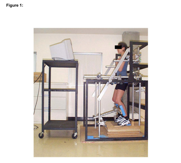
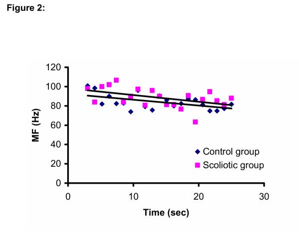
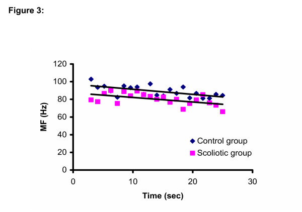
Methods: L5/S1 moments during isometric efforts in extension were measured in six subjects with idiopathic scoliosis and ten healthy controls. The subjects performed three 7 s ramp contractions ranging from 0 to 100% maximum voluntary contraction (MVC) and one 30 s sustained contraction at 75% MVC. Surface EMG activity was recorded bilaterally from the paraspinal muscles at L5, L3, L1 and T10. The slope of the EMG RMS/force (neuromuscular efficiency) and MF/force (muscle composition) relationships were computed during the ramp contractions while the slope of the EMG RMS/time and MF/time relationships (muscle fatigue) were computed during the sustained contraction. Comparisons were performed between the two groups and between the left and right sides for the EMG parameters.
Results: No significant group or side differences between the slopes of the different measures used were found at the level of the apex (around T10) of the major curve of the spine. However, a significant side difference was seen at a lower level (L3, p = 0.01) for the MF/time parameter.
Conclusion: The EMG parameters used in this study could not discriminate between the back muscles of scoliotic subjects and those of control subject regarding fiber type composition, neuromuscular efficiency and muscle fatigue at the level of the apex. The results of this pilot study indicate that compensatory strategies are potentially seen at lower level of the spine with these EMG parameters.
Figures
Similar articles
-
Asymmetry of paraspinal EMG-time characteristics in idiopathic scoliosis.J Spinal Disord. 1999 Feb;12(1):61-7. J Spinal Disord. 1999. PMID: 10078952
-
Between-days reliability of electromyographic measures of paraspinal muscle fatigue at 40, 50 and 60% levels of maximal voluntary contractile force.Clin Rehabil. 2002 Nov;16(7):761-71. doi: 10.1191/0269215502cr551oa. Clin Rehabil. 2002. PMID: 12428825
-
The electromyographic responses of paraspinal muscles during isokinetic exercise in adolescents with idiopathic scoliosis with a Cobb's angle less than fifty degrees.Chang Gung Med J. 2010 Sep-Oct;33(5):540-50. Chang Gung Med J. 2010. PMID: 20979705
-
The assessment of back muscle capacity using intermittent static contractions. Part I - Validity and reliability of electromyographic indices of fatigue.J Electromyogr Kinesiol. 2008 Dec;18(6):1006-19. doi: 10.1016/j.jelekin.2007.03.012. Epub 2007 Jul 20. J Electromyogr Kinesiol. 2008. PMID: 17643316 Review.
-
EMG assessment of back muscle function during cyclical lifting.J Electromyogr Kinesiol. 1998 Aug;8(4):233-45. doi: 10.1016/s1050-6411(98)00010-8. J Electromyogr Kinesiol. 1998. PMID: 9779397 Review.
Cited by
-
Electrophysiological and histological changes of paraspinal muscles in adolescent idiopathic scoliosis.Eur Spine J. 2016 Oct;25(10):3146-3153. doi: 10.1007/s00586-016-4628-8. Epub 2016 May 31. Eur Spine J. 2016. PMID: 27246349
-
Proposal of a new exercise protocol for idiopathic scoliosis: A preliminary study.Medicine (Baltimore). 2018 Dec;97(49):e13336. doi: 10.1097/MD.0000000000013336. Medicine (Baltimore). 2018. PMID: 30544395 Free PMC article.
-
Additive Manufacturing of Spinal Braces: Evaluation of Production Process and Postural Stability in Patients with Scoliosis.Materials (Basel). 2022 Sep 7;15(18):6221. doi: 10.3390/ma15186221. Materials (Basel). 2022. PMID: 36143533 Free PMC article.
-
Dynamical asymmetries in idiopathic scoliosis during forward and lateral initiation step.Eur Spine J. 2009 Feb;18(2):188-95. doi: 10.1007/s00586-008-0864-x. Epub 2009 Jan 8. Eur Spine J. 2009. PMID: 19130095 Free PMC article.
-
The Application of Surface Electromyography Technology in Evaluating Paraspinal Muscle Function.Diagnostics (Basel). 2024 May 24;14(11):1086. doi: 10.3390/diagnostics14111086. Diagnostics (Basel). 2024. PMID: 38893614 Free PMC article. Review.
References
-
- Salter RB. Textbook of disorders and injuries of the musculoskeletal system. second. Williams & Wilkins, Baltimore, Md; 1983.
-
- Yarom R, Robin GC. Studies on spinal and peripheral muscles from patients with scoliosis. Spine. 1979;4:12–21. - PubMed
-
- Kennelly KP, Stokes MJ. Pattern of asymmetry of paraspinal muscle size in adolescent idiopathic scoliosis examined by real-time ultrasound imaging. A preliminary study. Spine. 1993;18:913–917. - PubMed
-
- Gibson JNA, McMaster MJ, Scrimgeour CM, Stoward PJ, Rennie MJ. Rates of protein synthesis in paraspinal muscles: Lateral disparity in children with idiopathic scoliosis. Clinical Science. 1988;75:79–83. - PubMed
Publication types
MeSH terms
LinkOut - more resources
Full Text Sources
Other Literature Sources
Medical
Research Materials
Miscellaneous
