Selecting the best index for following the temporal evolution of apparent diffusion coefficient and diffusion anisotropy after hypoxic-ischemic white matter injury in neonates
- PMID: 15760851
- PMCID: PMC7976488
Selecting the best index for following the temporal evolution of apparent diffusion coefficient and diffusion anisotropy after hypoxic-ischemic white matter injury in neonates
Abstract
Background and purpose: Diffusion-weighted (DW) MR imaging is a useful technique for detecting ischemia. In adults and neonates, however, temporal changes on DW images after ischemia complicate interpretation. Our purpose was to investigate the temporal evolution of the apparent diffusion coefficient (ADC), diffusion-tensor (DT) imaging components, and anisotropy in neonatal brain after hypoxic-ischemic white matter injury and to determine which anisotropy index is preferable.
Methods: DT images were obtained with single-shot echo-planar imaging, by using pulsed field gradients in six directions. Sixteen volunteers and 10 term neonates with normal MR images were evaluated to obtain reference values. Among the anisotropy indexes of fractional anisotropy (FA), relative anisotropy (RA), volume ratio (VR), linear (CI) and planar (Cp) diffusion, and axial anisotropy (Am), simulations were performed to select the most appropriate indexes for clinical practice. The ADC, DT imaging components, and anisotropy were evaluated as a function of time after onset of symptoms in 11 neonates with hypoxic ischemia.
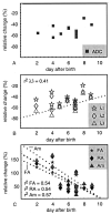
Results: In neonates, changes in lesions were characterized by a large decrease (40%) in all eigenvalues, with a stronger decrease in the direction perpendicular to the fibers, resulting in increased anisotropy indexes. The temporal evolution of the relative change in ADC did not show a significant trend (P >.05). The relative change in anisotropy decreased linearly with time (P <.05), with the strongest trend in anisotropy index Am.
Conclusion: In clinical practice, anisotropy indexes FA, RA and Am appear to be useful. Am is the best index to monitor anisotropy changes. DT imaging provides information about diffusion parallel and perpendicular to white matter fibers, which helps the interpretation of physiologic changes after hypoxic-ischemic injury.
Figures


References
-
- Moseley ME, Cohen Y, Mintorovitch J, et al. Early detection of regional cerebral ischemia in cats: comparison of diffusion- and T2-weighted MRI and spectroscopy. Magn Reson Med 1990;14:330–346 - PubMed
-
- Warach S, Chien D, Li W, Ronthal M, Edelman RR. Fast magnetic resonance diffusion-weighted imaging of acute human stroke. Neurology 1992;42:1717–1723 - PubMed
-
- Cowan FM, Pennock JM, Hanrahan JD, Manji KP, Edwards AD. Early detection of cerebral infarction and hypoxic ischemic encephalopathy in neonates using diffusion-weighted magnetic resonance imaging. Neuropediatrics 1994;25:172–175 - PubMed
-
- Inder T, Hüppi PS, Zientara GP, et al. Early detection of periventricular leukomalacia by diffusion-weighted magnetic resonance imaging techniques. J Pediatr 1999;134:631–634 - PubMed
MeSH terms
LinkOut - more resources
Full Text Sources
Miscellaneous
