

Late proton MR spectroscopy in children after traumatic brain injury: correlation with cognitive outcomes
- PMID: 15760852
- PMCID: PMC7976470
Late proton MR spectroscopy in children after traumatic brain injury: correlation with cognitive outcomes
Abstract
Background and purpose: Proton MR spectroscopy has demonstrated reduced levels of N-acetylaspartate (NAA) in normal-appearing occipital and frontal regions of patients with acute nonpenetrating traumatic brain injury (TBI). We studied the relationship of frontoparietal NAA, choline (Cho), and creatine (Cr) to test the hypothesis that reduction in NAA is predictive of cognitive outcome.
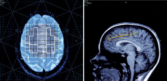
Methods: Proton spectra were collected by using conventional 2D chemical shift imaging in five healthy children and seven children (6 weeks to 3 years) with severe (n=4), moderate (n=2), or mild (n=1) TBI. Spectra in the anterior and posterior regions of the left and right frontoparietal areas were averaged for analysis by using LCModel, with a phantom-established basis function, for quantification of NAA, Cho, and Cr concentrations. Intellectual function, expressive language, and arithmetic capability were measured within 4 months of imaging.
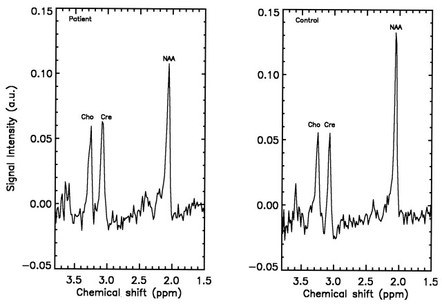
Results: NAA/Cho concentration was lower in TBI patients than in control subjects, but no group differences were present for Cho or Cr. Hemispheric levels for NAA, Cho, and Cr were higher on the left than on the right, but we found no effect of region and no interactions. Cognition was lower in the TBI group than the control group and correlated with NAA levels. Left frontal Cho was also correlated with arithmetic scores, whereas Cr was not significantly correlated.
Conclusion: NAA levels remain low after TBI and are related to cognitive function. Neurometabolite values are greater in the left frontoparietal region than in the right, and the left frontal Cho level is related to arithmetic ability.
Figures
Similar articles
-
Assessment of mitochondrial impairment in traumatic brain injury using high-resolution proton magnetic resonance spectroscopy.J Neurosurg. 2008 Jan;108(1):42-52. doi: 10.3171/JNS/2008/108/01/0042. J Neurosurg. 2008. PMID: 18173309 Clinical Trial.
-
Whole-brain proton MR spectroscopic imaging of mild-to-moderate traumatic brain injury and correlation with neuropsychological deficits.J Neurotrauma. 2010 Mar;27(3):483-96. doi: 10.1089/neu.2009.1159. J Neurotrauma. 2010. PMID: 20201668 Free PMC article.
-
Developmental delay in children: assessment with proton MR spectroscopy.AJNR Am J Neuroradiol. 2002 May;23(5):882-8. AJNR Am J Neuroradiol. 2002. PMID: 12006297 Free PMC article.
-
Magnetic Resonance Spectroscopy following Mild Traumatic Brain Injury: A Systematic Review and Meta-Analysis on the Potential to Detect Posttraumatic Neurodegeneration.Neurodegener Dis. 2020;20(1):2-11. doi: 10.1159/000508098. Epub 2020 Jul 1. Neurodegener Dis. 2020. PMID: 32610337
-
N-acetylaspartate as a marker of neuronal injury in neurodegenerative disease.Adv Exp Med Biol. 2006;576:241-62; discussion 361-3. doi: 10.1007/0-387-30172-0_17. Adv Exp Med Biol. 2006. PMID: 16802717 Free PMC article. Review. No abstract available.
Cited by
-
Magnetic resonance spectroscopy in pediatric neuroradiology: clinical and research applications.Pediatr Radiol. 2010 Jan;40(1):3-30. doi: 10.1007/s00247-009-1450-z. Epub 2009 Nov 24. Pediatr Radiol. 2010. PMID: 19937238
-
Emerging imaging tools for use with traumatic brain injury research.J Neurotrauma. 2012 Mar 1;29(4):654-71. doi: 10.1089/neu.2011.1906. Epub 2011 Oct 17. J Neurotrauma. 2012. PMID: 21787167 Free PMC article. Review.
-
Retinal metabolic changes in an experimental model of optic nerve transection by ex vivo 1H magnetic resonance spectroscopy.Neurochem Res. 2011 Dec;36(12):2427-33. doi: 10.1007/s11064-011-0570-7. Epub 2011 Aug 13. Neurochem Res. 2011. PMID: 21842271
-
Neuroimaging Correlates of Functional Outcome Following Pediatric TBI.Adv Neurobiol. 2024;42:33-84. doi: 10.1007/978-3-031-69832-3_3. Adv Neurobiol. 2024. PMID: 39432037 Review.
-
Challenges and opportunities for neuroimaging in young patients with traumatic brain injury: a coordinated effort towards advancing discovery from the ENIGMA pediatric moderate/severe TBI group.Brain Imaging Behav. 2021 Apr;15(2):555-575. doi: 10.1007/s11682-020-00363-x. Brain Imaging Behav. 2021. PMID: 32734437 Free PMC article.
References
-
- Tong K, Holshouser B, Shutter L, Chiou P, Herigauk G, Haacke E. High resolution susceptibility weighted imaging (SWI) improves detection of hemorrhagic lesions in adults with traumatic brain injury: correlation with severity of injury and outcome. Proceedings of ISMRM 10th Scientific Meeting. Berkeley: International Society for Magnetic Resonance in Medicine2002. :46 .
-
- Donders J, Strom D. Neurobehavioral recovery after pediatric head trauma: injury, pre- injury, and post-injury issues. J Head Trauma Rehabil 2000;15:792–803 - PubMed
-
- Brooks WM, Friedman SD, Gasparovic C. Magnetic resonance spectroscopy in traumatic brain injury. J Head Trauma Rehabil 2001;16:149–164 - PubMed
-
- Ashwal S, Holshouser BA, Shu SK, et al. Predictive value of proton magnetic resonance spectroscopy in pediatric closed head injury. Pediatr Neurol 2000;23:114–125 - PubMed
Publication types
MeSH terms
Substances
Grants and funding
LinkOut - more resources
Full Text Sources