Cortical lesions in multiple sclerosis: combined postmortem MR imaging and histopathology
- PMID: 15760868
- PMCID: PMC7976495
Cortical lesions in multiple sclerosis: combined postmortem MR imaging and histopathology
Abstract
Background and purpose: Cortical lesions constitute a substantial part of the total lesion load in multiple sclerosis (MS) brain. They have been related to neuropsychological deficits, epilepsy, and depression. However, the proportion of purely cortical lesions visible on MR images is unknown. The aim of this study was to determine the proportion of intracortical and mixed gray matter (GM)-white matter (WM) lesions that can be visualized with postmortem MR imaging.
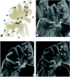
Methods: We studied 49 brain samples from nine cases of chronic MS. Tissue sections were matched to dual-echo T2-weighted spin-echo (T2SE) MR images. MS lesions were identified by means of myelin basic protein immunostaining, and lesions were classified as intracortical, mixed GM-WM, deep GM, or WM. Investigators blinded to the histopathologic results scored postmortem T2SE and 3D fluid-attenuated inversion recovery (FLAIR) images.
Results: Immunohistochemistry confirmed 70 WM, eight deep GM, 27 mixed GM-WM, and 63 purely cortical lesions. T2SE images depicted only 3% of the intracortical lesions, and 3D FLAIR imaging showed 5%. Mixed GM-WM lesions were most frequently detectable on T2SE and 3D FLAIR images (22% and 41%, respectively). T2SE imaging showed 13% of deep GM lesions versus 38% on 3D FLAIR. T2SE images depicted 63% of the WM lesions, whereas 3D FLAIR images depicted 71%. Even after side-by-side review of the MR imaging and histopathologic results, many of the intracortical lesions could not be identified retrospectively.
Conclusion: In contrast to WM lesions and mixed GM-WM lesions, intracortical lesions remain largely undetected with current MR imaging resolution.
Figures


References
-
- Dawson JW. The histology of multiple sclerosis. Trans R Soc Edinburgh 1916;50:517–740
-
- Lumsden CE. The neuropathology of multiple sclerosis. In: Vinken PJ, Bruin GW, ed. Handbook of Clinical Neurology. Amsterdam: Elsevier Science Publishers;1970. :217–309
-
- Kidd D, Barkhof F, McConnell R, Algra PR, Allen IV, Revesz T. Cortical lesions in multiple sclerosis. Brain 1999;122:17–26 - PubMed
-
- Peterson JW, Bo L, Mork S, Chang A, Trapp BD. Transected neurites, apoptotic neurons, and reduced inflammation in cortical multiple sclerosis lesions. Ann Neurol 2001;50:389–400 - PubMed
Publication types
MeSH terms
LinkOut - more resources
Full Text Sources
Medical
