Comparative Study
. 2005 May 1;564(Pt 3):791-801.
doi: 10.1113/jphysiol.2005.085704. Epub 2005 Mar 10.

Agonist activation of arachidonate-regulated Ca2+-selective (ARC) channels in murine parotid and pancreatic acinar cells

Affiliations
Comparative Study

Agonist activation of arachidonate-regulated Ca2+-selective (ARC) channels in murine parotid and pancreatic acinar cells

Olivier Mignen et al. J Physiol. .

Abstract

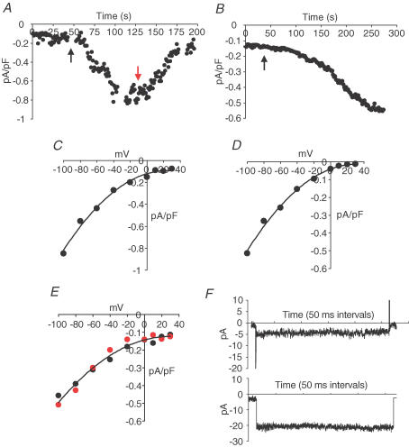
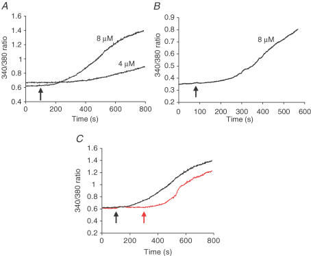
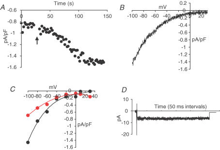
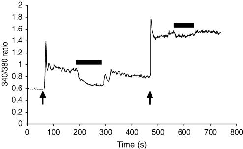
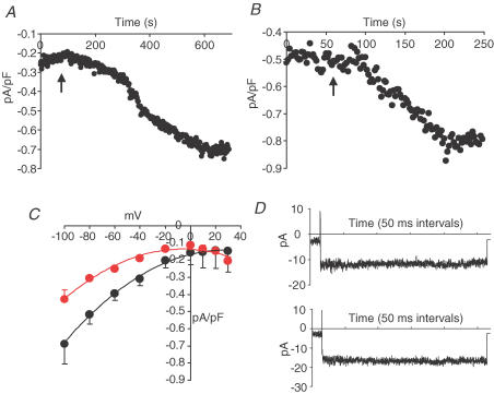
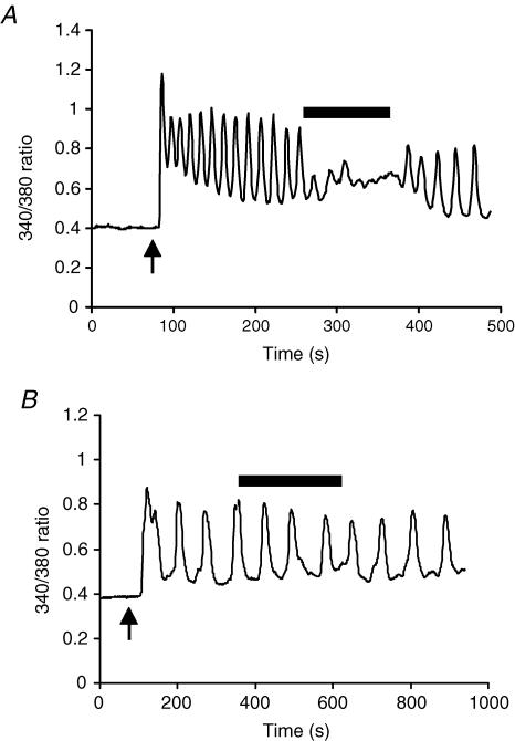
ARC channels (arachidonate-regulated Ca(2+)-selective channels) are a novel type of highly Ca(2+)-selective channel that are specifically activated by low concentrations of agonist-induced arachidonic acid. This activation occurs in the absence of any depletion of internal Ca(2+) stores (i.e. they are 'non-capacitative'). Previous studies in HEK293 cells have shown that these channels provide the predominant pathway for the entry of Ca(2+) seen at low agonist concentrations where oscillatory [Ca(2+)](i) signals are typically produced. In contrast, activation of the more widely studied store-operated Ca(2+) channels (e.g. CRAC channels) is only seen at higher agonist concentrations where sustained 'plateau-type'[Ca(2+)](i) responses are observed. We have now demonstrated the presence of ARC channels in both parotid and pancreatic acinar cells and shown that, again, they are specifically activated by the low concentrations of appropriate agonists (carbachol in the parotid, and both carbachol and cholecystokinin in the pancreas) that are associated with oscillatory [Ca(2+)](i) signals in these cells. Uncoupling the receptor-mediated activation of cytosolic phospholipase A(2) (cPLA(2)) with isotetrandrine reduces the activation of the ARC channels by carbachol and, correspondingly, markedly inhibits the [Ca(2+)](i) signals induced by low carbachol concentrations, whilst those signals seen at high agonist concentrations are essentially unaffected. Interestingly, in the pancreatic acinar cells, activation by cholecystokinin induces a current through the ARC channels that is only approximately 60% of that seen with carbachol. This is consistent with previous reports indicating that carbachol-induced [Ca(2+)](i) signals in these cells are much more dependent on Ca(2+) entry than are the cholecystokinin-induced responses.


Figures

Figure 1
Figure 1. Arachidonic acid-activated currents in parotid and pancreatic acinar cells
A and B, representative traces showing the effect of exogenous arachidonic acid (8 μm, added at black arrow) on currents measured at −80 mV in single isolated parotid acinar cells (A), and isolated pancreatic acinar cells (B). In A, La3+ (50 μm) was added at the red arrow. C and D, representative current–voltage relationships of the currents activated by addition of arachidonic acid (8 μm) in parotid (C) and pancreatic acinar cells (D). E, comparison of the arachidonic acid-activated currents in pancreatic acinar cells measured in normal external solution (black symbols), and that measured in a solution in which external Na+ was replaced with the impermeant cation NMDG+ (red symbols). F, representative currents recorded during 250 ms pulses to −80 mV from a holding potential of 0 mV in isolated parotid acinar cells (top trace) and pancreatic acinar cells (lower trace) following activation with arachidonic acid (8 μm). Capacity transients were corrected as described in Methods.
Figure 2
Figure 2. Effect of arachidonic acid on [Ca2+]i in parotid and pancreatic acinar cells
A and B, representative traces showing the effect of adding arachidonic acid at the indicated concentrations (black arrow) on [Ca2+]i measured as the fluorescence ratio (F340/380) of intracellularly loaded fura-2 in isolated parotid acinar cells (A), and isolated pancreatic acinar cells (B). C, inhibition of the arachidonic acid-induced increase in [Ca2+]i in parotid cells by La3+. Arachidonic acid (8 μm) was added (black arrow) either in the absence (black trace) or presence (red trace) of 50 μm La3+. The La3+ was subsequently removed (red arrow).
Figure 3
Figure 3. Activation of ARC channels by low concentrations of carbachol in parotid acinar cells
A, representative trace showing the effect of carbachol (250 nm, added at arrow) on currents measured at −80 mV in single isolated parotid acinar cells. B, representative current–voltage relationship of the current activated by 250 nm carbachol in parotid acinar cells. C, representative current–voltage relationships of the currents activated by 250 nm carbachol in parotid acinar cells in the presence (red trace) and absence (black trace) of isotetrandrine (10 μm). D, representative current recorded during a 250 ms pulse to −80 mV from a holding potential of 0 mV in an isolated parotid acinar cell following activation with 250 nm carbachol measured in the presence of 10 μm isotetrandrine. Capacity transients were corrected as described in Methods.
Figure 4
Figure 4. Effect of isotetrandrine on the [Ca2+]i signals activated by low and high carbachol concentrations in isolated parotid acinar cells
[Ca2+]i was measured as the F340/380 ratio of intracellularly loaded fura-2 as described. At the first arrow, carbachol (300 nm) was added; at the second arrow the carbachol concentration was increased to 10 μm. The black bars indicate when isotetrandrine (10 μm) was present.
Figure 5
Figure 5. Activation of ARC channels by carbachol and cholecystokinin in pancreatic acinar cells
A and B, representative traces showing the effect of carbachol (200 nm) and CCK-8 (20 pm), respectively, on currents measured at −80 mV in single isolated pancreatic acinar cells. The agonists were added at the arrow in each case. C, mean (± s.e.m.) current–voltage relationships of the currents activated by carbachol (200 nm, black circles), and CCK-8 (20 pm, red circles) in isolated pancreatic acinar cells. D, representative currents recorded during 250 ms pulses to −80 mV from a holding potential of 0 mV in isolated pancreatic acinar cells in the presence of carbachol (200 nm, top trace) and CCK-8 (20 pm, lower trace). Capacity transients were corrected as described in Methods.
Figure 6
Figure 6. Effect of isotetrandrine on the [Ca2+]i signals activated by carbachol and cholecystokinin in pancreatic acinar cells
Changes in [Ca2+]i in isolated pancreatic acinar cells, measured as F340/380 of intracellularly loaded fura-2, following addition of 200 nm carbachol (A), or 20 pm CCK-8 (B). In each case, the agonists were added at the arrow. Subsequently, isotetrandrine (10 μm) was added as indicated by the black bar. Notice the different time scales of the responses.

Similar articles

Cited by

References

    1. Akiba S, Kato E, Sato T, Fujii T. Biscoclaurine alkaloids inhibit receptor-mediated phospholipase A2 activation probably through uncoupling of a GTP-binding protein from the enzyme in rat peritoneal mast cells. Biochem Pharmacol. 1992;44:45–50. 10.1016/0006-2952(92)90036-I. - DOI - PubMed
    1. Begenisich T, Nakamoto T, Ovitt CE, Nehrke K, Brugnara C, Alper SL, Melvin JE. Physiological roles of the intermediate conductance, Ca2+-activated potassium channel Kcnn4. J Biol Chem. 2004;279:47681–47687. - PubMed
    1. Bers DM, Patton CW, Nuccitelli R. A practical guide to the preparation of Ca2+ buffers. Meths Cell Biol. 1994;40:1–29. - PubMed
    1. Bird GS, Putney JW., Jr Capacitative calcium entry supports calcium oscillations in human embryonic kidney cells. J Physiol. 2005;562:697–706. - PMC - PubMed
    1. Broad LM, Cannon TR, Taylor CW. A non-capacitative pathway activated by arachidonic acid is the major Ca2+ entry mechanism in rat A7r5 smooth muscle cells stimulated with low concentrations of vasopressin. J Physiol. 1999;517:121–134. - PMC - PubMed

Publication types

LinkOut - more resources