. 2005 May 1;564(Pt 3):907-21.
doi: 10.1113/jphysiol.2005.084780. Epub 2005 Mar 10.

Protein kinase A-dependent enhanced NMDA receptor function in pain-related synaptic plasticity in rat amygdala neurones

Affiliations

Protein kinase A-dependent enhanced NMDA receptor function in pain-related synaptic plasticity in rat amygdala neurones

Gary C Bird et al. J Physiol. .

Abstract

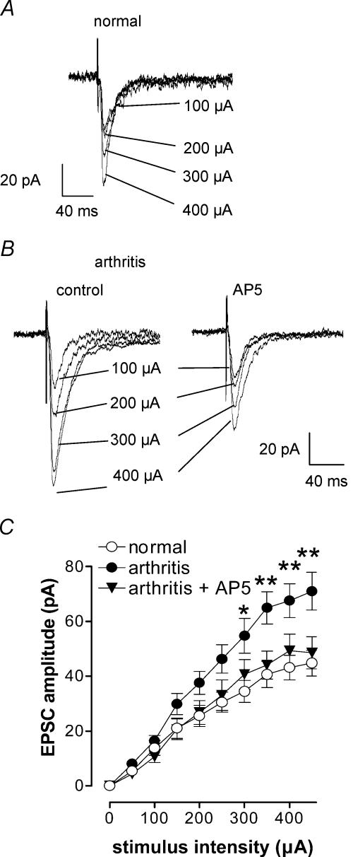
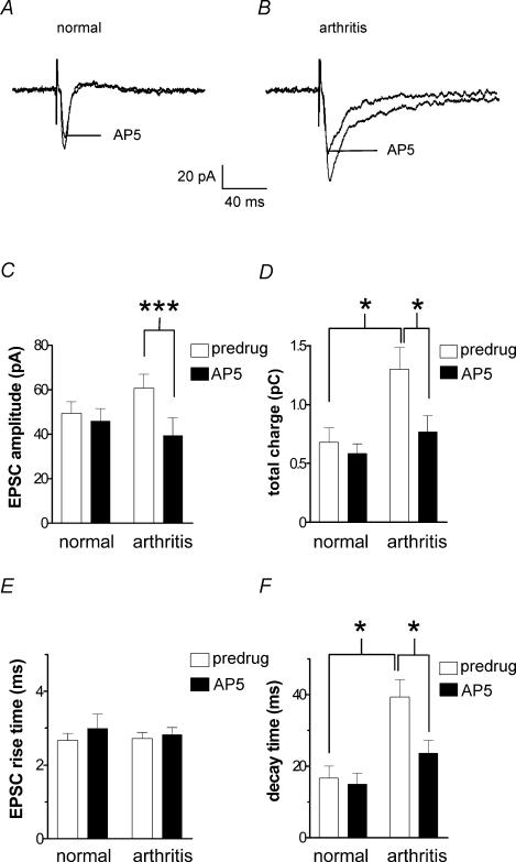
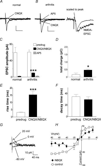
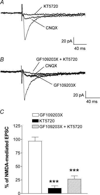
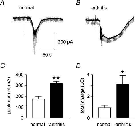
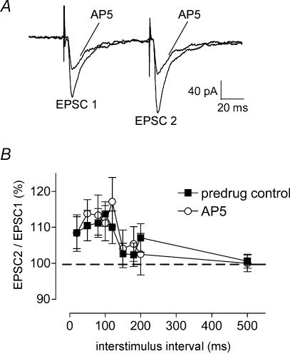
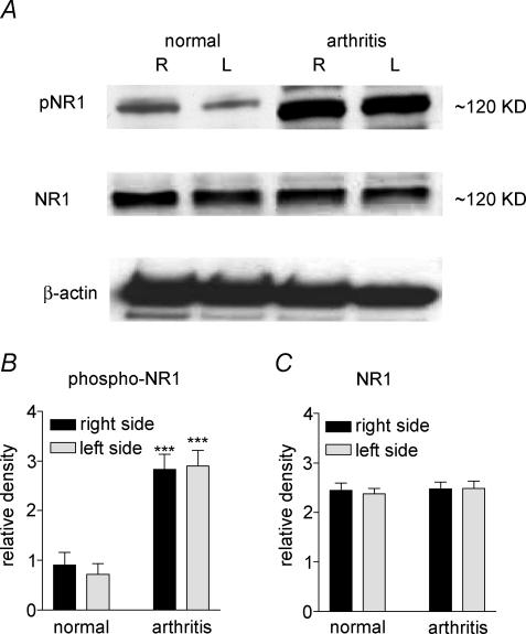
Mechanisms of pain-related plasticity in the amygdala, a key player in emotionality, were studied at the cellular and molecular levels in a model of arthritic pain. The influence of the arthritis pain state induced in vivo on synaptic transmission and N-methyl-d-aspartate (NMDA) receptor function was examined in vitro using whole-cell voltage-clamp recordings of neurones in the latero-capsular part of the central nucleus of the amygdala (CeA), which is now defined as the 'nociceptive amygdala'. Synaptic transmission was evoked by electrical stimulation of afferents from the pontine parabrachial area (part of the spino-parabrachio-amygdaloid pain pathway) in brain slices from control rats and from arthritic rats. This study shows that pain-related synaptic plasticity is accompanied by protein kinase A (PKA)-mediated enhanced NMDA-receptor function and increased phosphorylation of NMDA-receptor 1 (NR1) subunits. Synaptic plasticity in the arthritis pain model, but not normal synaptic transmission in control neurones, was inhibited by a selective NMDA receptor antagonist. Accordingly, an NMDA receptor-mediated synaptic component was recorded in neurones from arthritic animals, but not in control neurones, and was blocked by inhibition of PKA but not protein kinase C (PKC). Exogenous NMDA evoked a larger inward current in neurones from arthritic animals than in control neurones, indicating a postsynaptic effect. Paired-pulse facilitation, a measure of presynaptic mechanisms, was not affected by an NMDA-receptor antagonist. Increased levels of phosphorylated NR1 protein, but not of total NR1, were measured in the CeA of arthritic rats compared to controls. Our results suggest that pain-related synaptic plasticity in the amygdala involves a critical switch of postsynaptic NMDA receptor function through PKA-dependent NR1 phosphorylation.


Figures

Figure 1
Figure 1. Synaptic plasticity in CeA neurones in the arthritis pain model
Whole-cell voltage-clamp recordings were made from neurones in the latero-capsular part of the CeA in brain slices from normal rats and from arthritic rats (6–8 h postinduction). Monosynaptic excitatory postsynaptic currents (EPSCs) were evoked at the nociceptive parabrachial (PB)-CeA synapse (Bourgeais et al. 2001; Gauriau & Bernard, 2002; Neugebauer et al. 2004). A and B, individual traces recorded in one CeA neurone from a normal rat (A) and another CeA neurone from an arthritic rat (B) in control and in the presence of an NMDA receptor antagonist (AP5, 50 μm). Traces represent the mean of eight to 12 trials and stimulus artifacts have been truncated. C, input–output curves were generated by plotting peak EPSC amplitude as a function of increasing stimulus intensity (see Methods). Input–output relationships of neurones from arthritic animals (n = 51) were significantly different (P < 0.0001, F1,780= 40.18, two-way ANOVA) from those of control neurones (n = 29), suggesting enhanced synaptic transmission in the CeA in the arthritis pain model. In arthritis, the presence of AP5 significantly changed the input–output relationship (P < 0.0001, F9,590= 16.19, two-way ANOVA, n = 10). Neurones were voltage-clamped at −60 mV *P < 0.05, **P < 0.01, Bonferroni post hoc test.
Figure 2
Figure 2. Inhibition of synaptic transmission by an NMDA receptor antagonist (AP5) is enhanced in the arthritis pain model
A and B, monosynaptic EPSCs recorded in a CeA neurone from a normal rat (A) and in another neurone from an arthritic rat (B). AP5 (50 μm) inhibited synaptic transmission in arthritis but not under normal conditions. C and D, averaged raw data show that AP5 inhibited peak EPSC amplitude (C) and total charge (area under the curve, D) of monosynaptic EPSCs in arthritis (n = 11 neurones) but not in CeA neurones from normal animals (n = 12). Increase of the total charge of EPSC in the arthritis pain model suggests increased synaptic strength. E and F, differential effects of AP5 (50 μm) on the 10–90% rise time (E) and decay time constant (t, F) in neurones from arthritic rats (n = 11 neurones) and from normal animals (n = 10 neurones). AP5 shortened the increased decay time of monosynaptic EPSCs in the arthritis pain model. Monosynaptic EPSCs were evoked at the PB-CeA synapse using a stimulus intensity that generated 80% of the maximum EPSC amplitude. Neurones were voltage-clamped at −60 mV. *P < 0.05, ***P < 0.001, paired and unpaired t tests.
Figure 3
Figure 3. Pharmacological isolation of an NMDA receptor-mediated EPSC in synaptic plasticity in the arthritis pain model
A and B, monosynaptic EPSCs recorded in a CeA neurone from a normal rat (A) and in another neurone from an arthritic rat (B). CNQX (30 μm) blocked synaptic transmission in the control neurone from a normal animal, but a resistant component remained in arthritis. Addition of AP5 (50 μm) blocked the CNQX-resistant component in arthritis. Traces to the right of B are scaled to the peak to illustrate the slow decay time of the NMDA EPSC. Scale bars in A and B are 20 pA and 40 ms, respectively. C, averaged raw data show a significant CNQX/NBQX-resistant EPSC component (measured as peak amplitude) in arthritis (n = 31; P < 0.001, paired t test), but not in control neurones from normal rats (n = 9). D, averaged raw data show the total charge of the NMDA receptor-mediated EPSC component is significantly increased in arthritis; recordings were made in the presence of CNQX/NBQX (P < 0.05, unpaired t test). E and F, averaged data show that the 10–90% rise time of the CNQX/NBQX-resistant EPSC component is significantly increased (E; P < 0.001, paired t test) compared to the predrug compound EPSC, but decay time is not affected (F). G and H, voltage-dependence of the NMDA-mediated EPSC. G, monosynaptic EPSCs recorded in the presence of NBQX (10 μm) at different holding potentials in an individual CeA neurone in a slice from an arthritic rat. H, current–voltage relationship of the NMDA receptor-mediated EPSC recorded in the presence of NBQX (10 μm) compared to the compound EPSC (predrug control) in CeA neurones (n = 3) from arthritic animals. The change in slope is consistent with the removal of a non-NMDA receptor-mediated EPSC component. *P < 0.05, ***P < 0.001.
Figure 4
Figure 4. A PKA inhibitor (KT5720), but not a PKC inhibitor (GF109203X), blocks the NMDA receptor-mediated synaptic plasticity
A, monosynaptic EPSCs recorded in a CeA neurone from an arthritic rat are only partially inhibited by CNQX (10 μm). This NMDA receptor-mediated component is completely blocked by KT5720 (1 μm). B, in another CeA neurone, application of GF109203X (1 μm) had no effect on the CNQX/NBQX-resistant NMDA receptor-mediated EPSC, but addition of KT5720 (1 μm) completely blocked the response. C, averaged normalized data show a significant inhibition of the CNQX/NBQX-resistant NMDA receptor-mediated synaptic component by KT5720 (n = 7) and by co-application of GF109203X and KT5720 (n = 7), but not by GF109203X alone (n = 8). Monosynaptic EPSCs were evoked at the PB-CeA synapse using a stimulus intensity that generated 80% of the maximum EPSC amplitude. Each trace in A and B represents the average of 10–12 EPSCs. Neurones were voltage-clamped at −60 mV. ***P < 0.001, unpaired t test (compared to predrug controls in the presence of CNQX).
Figure 5
Figure 5. Direct membrane effect of NMDA is increased in CeA neurones in the arthritis pain model
A and B, current traces show that NMDA (2 mm) evokes a larger inward current in a neurone recorded in a brain slice from an arthritic animal (B) than in another neurone from a normal animal (A). In each experiment one microdrop (10 μl) of NMDA was applied to the recording chamber using a pipette (see artifact) as described in the Methods. Hyperpolarizing voltage steps producing inward currents (see downward deflections) were used for the continuous monitoring of membrane conductance changes. Sodium spike currents appeared around the peak of the NMDA-evoked membrane current. C and D, averaged data show that significantly larger inward currents were evoked by NMDA in arthritis (n = 7 neurones) than under normal conditions (n = 4 neurones) both in terms of peak current amplitude (C) and total charge (area under the curve, D). Neurones were voltage-clamped at −60 mV. *P < 0.05, **P < 0.01, unpaired t test.
Figure 6
Figure 6. Paired-pulse facilitation (PPF) is not altered by block of NMDA receptors
The effect of AP5 on PPF was studied in CeA neurones in brain slices from arthritic rats. PPF, a measure of presynaptic transmitter release mechanisms, was evoked by electrical stimulation of the PB-CeA synapse at progressively increasing interstimulus intervals. A, pairs of monosynaptic EPSCs were recorded in one CeA neurone before and during application of AP5 (50 μm). Each trace is the average of 10–12 EPSCs. B, averaged normalized data show that PPF in the presence of AP5 (n = 12 neurones) was not significantly different than PPF before drug application (predrug control; n = 14 neurones). Peak amplitudes were measured as the difference between the current level before the stimulus artifact (see A) and the peak of the EPSC. PPF was calculated as EPSC2/EPSC1 × 100. Neurones were voltage-clamped at −60 mV. The lack of changes of PPF during block of endogenous NMDA-receptors argues against the involvement of any presynaptic NMDA receptors.
Figure 7
Figure 7. Increased phospho-NR1, but not total NR1, expression in the CeA in the arthritis pain model
A, immunoblots of phosphorylated NR1 (pNR1) and total NR1 in the CeA of the right (R, contralateral to the arthritis) and left (L, ipsilateral) brain hemispheres of normal and arthritic rats (6–8 h postinduction) are shown compared to a β-actin standard. B and C, averaged data of immunoblot densitometry show that the relative density of phospho-NR1 protein (B) is significantly increased in the right and left CeA of arthritic rats (n = 6) compared to normal rats (n = 6; P < 0.001, unpaired t test). There was no significant difference of total NR1 protein expression (C) in normal (n = 6) and arthritic (n = 6) rats.

References

    1. Becerra LR, Breiter HC, Stojanovic M, Fishman S, Edwards A, Comite AR, Gonzalez RG. Human brain activation under controlled thermal stimulation and habituation of noxious heat: an fMRI study. Magn Reson Med. 1999;41:1044–1057. - PubMed
    1. Bingel U, Quante M, Knab R, Bromm B, Weiller C, Buchel C. Subcortical structures involved in pain processing: evidence from single-trial fMRI. Pain. 2002;99:313–321. - PubMed
    1. Bornhovd K, Quante M, Glauche V, Bromm B, Weiller C, Buchel C. Painful stimuli evoke different stimulus-response functions in the amygdala, prefrontal, insula and somatosensory cortex: a single-trial fMRI study. Brain. 2002;125:1326–1336. - PubMed
    1. Bourgeais L, Gauriau C, Bernard J-F. Projections from the nociceptive area of the central nucleus of the amygdala to the forebrain: a PHA-L study in the rat. Eur J Neurosci. 2001;14:229–255. - PubMed
    1. Burnashev N, Schoepfer R, Monyer H, Ruppersberg JP, Gunther W, Seeburg PH, Sakmann B. Control by asparagine residues of calcium permeability and magnesium blockade in the NMDA receptor. Science. 1992;257:1415–1419. - PubMed

Publication types

LinkOut - more resources