. 2005 Jun 1;565(Pt 2):517-35.
doi: 10.1113/jphysiol.2005.083436. Epub 2005 Mar 10.

Inner and outer retinal pathways both contribute to surround inhibition of salamander ganglion cells

Affiliations

Inner and outer retinal pathways both contribute to surround inhibition of salamander ganglion cells

Tomomi Ichinose et al. J Physiol. .

Abstract

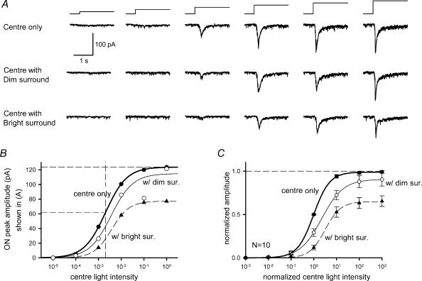
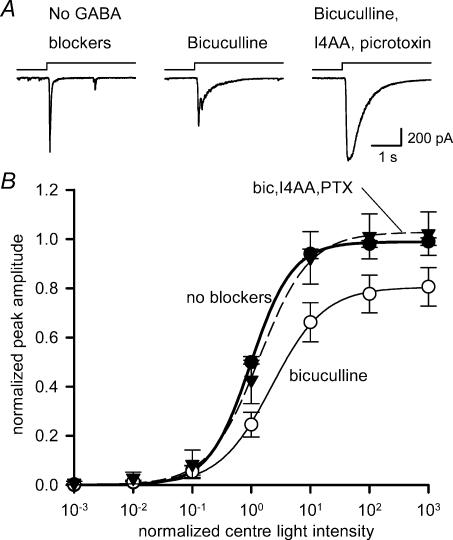
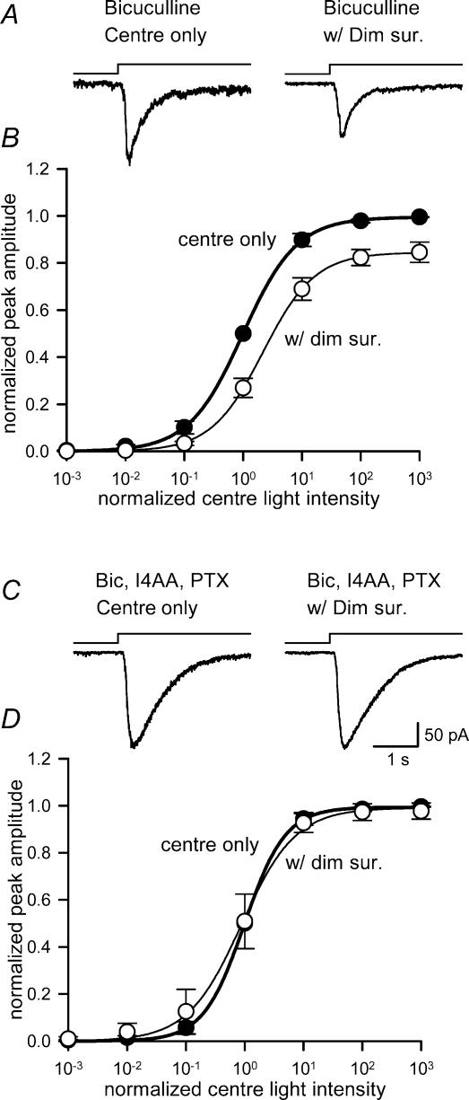
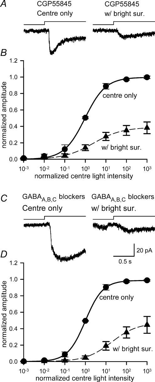
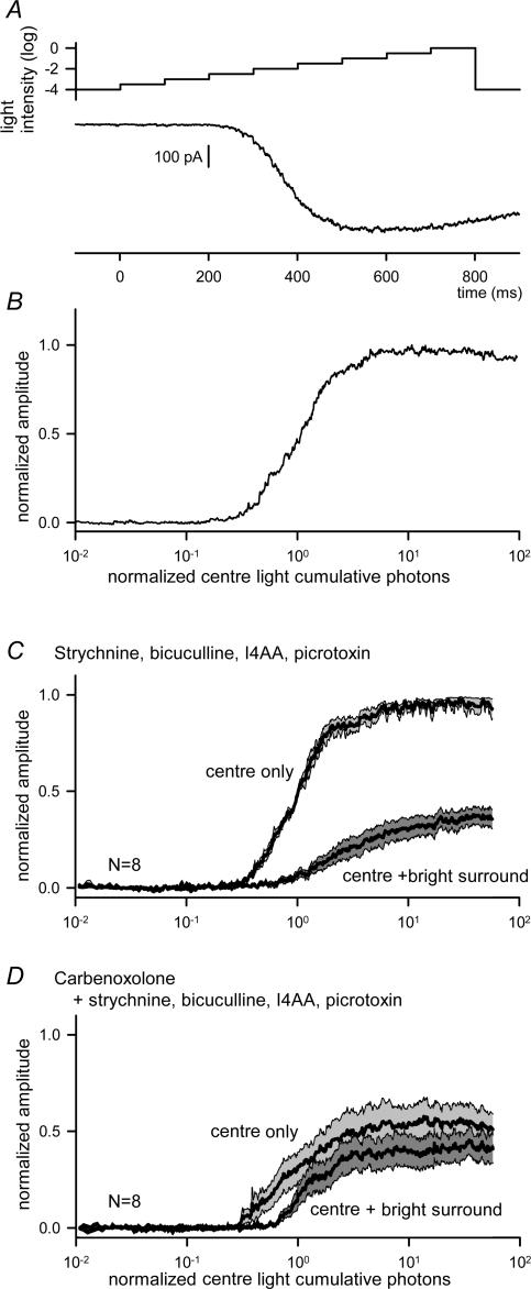
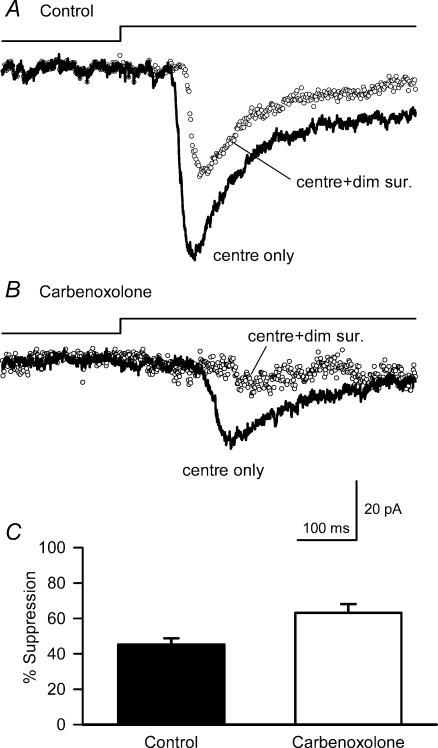
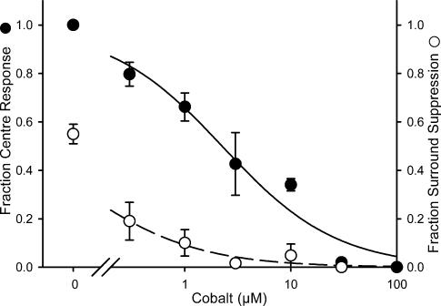
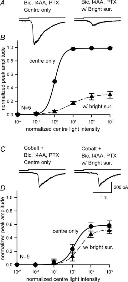
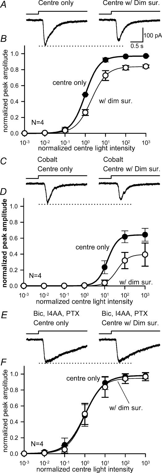
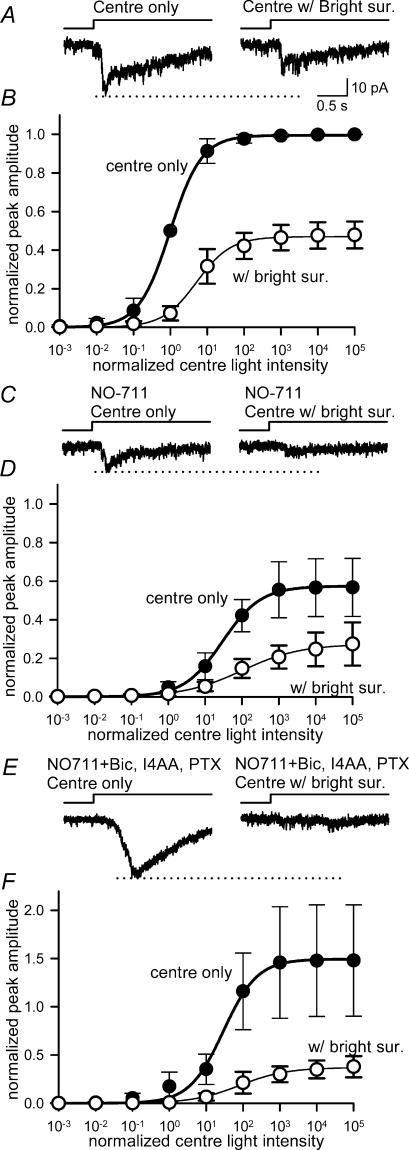
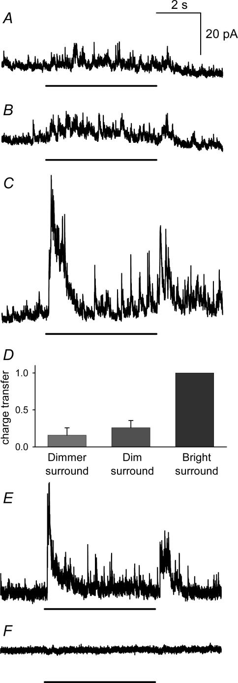
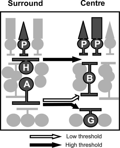
Illumination of the receptive-field surround reduces the sensitivity of a retinal ganglion cell to centre illumination. The steady, antagonistic receptive-field surround of retinal ganglion cells is classically attributed to the signalling of horizontal cells in the outer plexiform layer (OPL). However, amacrine cell signalling in the inner plexiform layer (IPL) also contributes to the steady receptive-field surround of the ganglion cell. We examined the contributions of these two forms of presynaptic lateral inhibition to ganglion cell light sensitivity by measuring the effects of surround illumination on EPSCs evoked by centre illumination. GABA(C) receptor antagonists reduced inhibition attributed to dim surround illumination, suggesting that this inhibition was mediated by signalling to bipolar cell axon terminals. Brighter surround illumination further reduced the light sensitivity of the ganglion cell. The bright surround effects on the EPSCs were insensitive to GABA receptor blockers. Perturbing outer retinal signalling with either carbenoxolone or cobalt blocked the effects of the bright surround illumination, but not the effects of dim surround illumination. We found that the light sensitivities of presynaptic, inhibitory pathways in the IPL and OPL were different. GABA(C) receptor blockers reduced dim surround inhibition, suggesting it was mediated in the IPL. By contrast, carbenoxolone and cobalt reduced bright surround, suggesting it was mediated by horizontal cells in the OPL. Direct amacrine cell input to ganglion cells, mediated by GABA(A) receptors, comprised another surround pathway that was most effectively activated by bright illumination. Our results suggest that surround activation of lateral pathways in the IPL and OPL differently modulate the sensitivity of the ganglion cell to centre illumination.


Figures

Figure 1
Figure 1. Surround illumination reduced ganglion cell EPSCs evoked by receptive-field centre illumination
A, EPSCs evoked by centre illumination (L-EPSCs) of increasing intensity (10−5–10° relative intensity, from left to right) in a ganglion cell voltage-clamped at −58.7 mV (ECl) (upper traces). Dim surround illumination (10−3 relative intensity) attenuated the L-EPSCs (middle traces). Bright surround illumination (10−1 relative intensity) reduced L-EPSCs further (lower traces). The time course and relative intensity of centre illumination is indicated by the stimulus traces above the light responses here and in subsequent figures. B, the peak amplitude of L-EPSCs (•), or with dim surround (○), or with bright surround (▴) in A was plotted as a function of centre light intensity. Each light intensity–response curve was fitted by the Hill equation (see Methods). The dashed horizontal lines indicate maximum amplitude (top) and half-maximum amplitude (middle) of L-EPSCs. The dashed vertical line indicates the light intensity at half-maximum of the centre-evoked EPSCs (L50). C, in this, and all of the succeeding intensity–response curves, the control centre illumination intensity–response curves were normalized to both the maximum amplitude of L-EPSCs, and to the light intensity at half-maximum amplitude of L-EPSCs. Curves measured in the presence of surround illumination were shifted relative to the control curve. The average maximum amplitude and L50 for the centre light were 0.99 ± 0.02 and 1.00 ± 0.0, respectively (n = 10). Dim surround illumination shifted the L50 to higher intensities (P < 0.01, n = 5), but did not significantly reduce the maximum amplitude (P = 0.19). Bright surround illumination shifted L50 values further (P < 0.01, n = 7) and reduced the maximum amplitudes (P < 0.01).
Figure 2
Figure 2. Effects of GABAA and GABAC receptor antagonists on EPSCs evoked by centre illumination
A, the amplitude of a ganglion cell EPSC evoked by centre illumination (left) was reduced by the GABAA receptor blocker bicuculline (200 μm) (middle). The suppressive effects of bicuculline were reversed by the addition of GABAC blockers, 200 μm picrotoxin and 20 μm I4AA (right). B, intensity–response curves for the centre-evoked EPSCs in the absence (•) or presence of GABA receptor blockers. Bicuculline (○) shifted the curve to the right (P < 0.05; n = 8) and reduced the maximum amplitude (P < 0.05). GABAC receptor blockers (▾) reversed the effects of bicuculline and shifted the light–response curve back to control levels (L50, P = 0.07; max, P = 0.35; n = 10).
Figure 3
Figure 3. GABAC receptor antagonists blocked the reduction in ganglion cell light sensitivity by dim surround illumination
A, a ganglion cell L-EPSC in the presence of bicuculline (left), was reduced by dim surround illumination (right). B, in the presence of bicuculline, the intensity–response curves for centre illumination (•) were still shifted to the right by the dim surround (○), indicating that bicuculline did not reverse the inhibition attributed to dim surround illumination (L50, P < 0.05; max, P < 0.05; n = 4). C, L-EPSCs recorded in the presence of bicuculline and the GABAC receptor antagonists, I4AA and picrotoxin (left), were unaffected by dim surround illumination (right). D, in the presence of GABAA and GABAC receptor antagonists, the intensity–response curves for centre plus surround (○) were similar to those for centre illumination alone (•) (L50, P = 0.40; max, P = 0.22; n = 5).
Figure 4
Figure 4. GABAA or GABAC receptor antagonists did not block the effects of bright surround illumination
A, L-EPSCs measured in the presence of bicuculline (left) were still strongly suppressed by bright surround illumination (right). B, in the presence of bicuculline, the intensity–response curves for centre illumination (•) were still shifted by bright surround illumination (▴), indicating that bicuculline did not block the inhibition evoked by bright surround illumination (L50, P < 0.05; max, P < 0.01; n = 6). C, L-EPSCs measured after the GABAC receptor blockers, I4AA and picrotoxin, were applied with bicuculline were still reduced by bright surround illumination. D, in the presence of GABAA and GABAC receptor antagonists, the centre intensity–response curve (•) was still shifted to the right and compressed by bright surrounds (▴) (L50, P < 0.05; max, P < 0.01; n = 7), indicating that these receptors did not mediate the effects of bright surround illumination.
Figure 5
Figure 5. Bright surround inhibition was not mediated by GABAB receptors
A, L-EPSCs measured in the presence of the GABAB receptor antagonist, CGP55845 (left) were still attenuated by bright surround illumination (right). B, the centre intensity–response curve (•) was still shifted to the right and compressed by bright surround light (▴) (L50, P < 0.05; max, P < 0.01; n = 5). C, the L-EPSC (left) measured in the presence of bicuculline, CGP55845, I4AA and picrotoxin was still attenuated by bright surround illumination (right). D, the centre light, intensity–response curve (•) was compressed and shifted to right with bright surround light (▴) (L50, P < 0.05; max, P < 0.01; n = 5).
Figure 6
Figure 6. Carbenoxolone reduced the inhibition by bright surrounds
A, ramp-light stimulation protocol consisted of 100 ms and 0.5 log intensity steps from −4 log to 0 log attenuation (upper trace) and a representative EPSC in response to the ramp light stimulation in the presence of strychnine, bicuculline, I4AA and picrotoxin (lower trace). B, the cumulative photon–response relationship obtained by re-plotting the EPSC in A. C, the cumulative photon–response relations (mean ± s.e.m.) from eight ganglion cells obtained in the absence or the presence of bright surround light stimulation (‘centre only’ and ‘centre + bright surround’, respectively). Bright surround illumination shifted the curve to the right and compressed it (L50, P = 0.01; max, P < 0.01; n = 8). D, in the presence of carbenoxolone, the cumulative photon–response relation was compressed (L50, P = 0.16; max, P < 0.01; n = 8), while surround inhibition was eliminated (L50, P = 0.11 versus‘centre only’; max, P = 0.12 versus‘centre only’; n = 8).
Figure 7
Figure 7. Carbenoxolone did not affect dim surround inhibition
A, L-EPSCs in the presence of strychnine, but in the absence of GABAA and GABAC receptor antagonists (thick line), was suppressed by 43% by dim surround illumination (○). B, the L-EPSC was still suppressed (by 43%) by dim surround illumination when carbenoxolone (100 μm) was present, although it reduced the EPSC evoked by the centre illumination (see text). C, bar graph summarizing the dim surround illumination results. The suppression of L-EPSCs by dim surround illumination (45.3 ± 3.5%, filled bar) was not reduced by carbenoxolone (63.2 ± 5.0%, n = 7, open bar).
Figure 8
Figure 8. Low concentrations of cobalt attenuated the bright surround inhibition as well as EPSCs evoked by centre illumination
L-EPSCs were recorded in the presence or absence of bright surround illumination. EPSCs evoked by centre light alone were attenuated by cobalt at 0.3 μm (•), and were abolished at 50 μm. The suppressive effect of bright surround inhibition was also blocked with cobalt at 0.3 μm (○), and was mostly eliminated at 3 μm. Each point shows the mean (± s.e.m.) of responses from different neurones (n = 2–3).
Figure 9
Figure 9. Cobalt blocked the reduction in ganglion cell light sensitivity by bright surround illumination
A, L-EPSCs in the presence of bicuculline, I4AA and picrotoxin (left), were reduced by bright surround illumination (right). B, in the presence of GABAA and GABAC receptor antagonists, the intensity–response curves for centre illumination (•) were still shifted to the right by the bright surround (▴) (L50, P < 0.01; max, P < 0.01; n = 5). C, L-EPSCs recorded in the presence of 3–10 μm cobalt as well as GABAA and GABAC receptor antagonists (left) were unaffected by bright surround illumination (right). D, in the presence of 3–10 μm cobalt, as well as GABAA and GABAC receptor antagonists, the intensity–response curve for centre plus surround illumination (▴) was similar to the curve for centre illumination alone (•) (L50, P = 0.14; max, P = 0.08; n = 5).
Figure 10
Figure 10. Cobalt did not block the effects of dim surround illumination
A, ganglion cell EPSCs evoked by centre illumination (left), were reduced by dim surround illumination (right) in the control Ringer solution. B, the intensity–response curves for centre illumination (•) were shifted to the right and compressed by the dim surround illumination (○) (L50, P < 0.05; max, P < 0.01; n = 4). C, 10 μm cobalt attenuated centre-evoked EPSCs (left). The EPSCs were still suppressed by dim surround illumination (right). D, in the presence of 10 μm cobalt, the intensity–response curves for centre illumination (•) were still shifted by dim surround illumination (○), indicating that cobalt did not affect the inhibition evoked by dim surround illumination (L50, P < 0.05; max, P < 0.05; n = 4). E, after cobalt was washed out, L-EPSCs were recorded in the presence of GABAA and GABAC receptor antagonists (left). Dim surround illumination did not attenuate the L-EPSCs (right). F, in the presence of GABAA and GABAC receptor antagonists, the centre intensity–response curve (•) was similar to the curve for centre with dim surround illumination (○) (L50, P = 0.13; max, P = 0.37; n = 4), indicating that dim surround inhibition was mediated by GABA receptors, but not by cobalt-sensitive pathways.
Figure 11
Figure 11. Bright surround inhibition did not utilize a presynaptic, IPL pathway
A, L-EPSCs recorded in control solution (left), were attenuated by bright surround illumination (right). B, the centre intensity–response curve (•) was shifted to the right and compressed by bright surround light (○) (L50, P < 0.05; max, P < 0.05; n = 3). C, NO-711 (3 μm) reduced L-EPSCs (left), which was further reduced by bright surround illumination (right). D, the intensity–response relationships of L-EPSCs in the presence of NO-711, with and without bright surround illumination. Bright surrounds still shifted the curve to the right (L50, P < 0.05; max, P < 0.09; n = 3). E, subsequent application of ionotropic GABA receptor blockers, 200 μm bicuculline, 50 μm I4AA and 200 μm picrotoxin, reversed the effect of NO-711 on centre-evoked L-EPSCs (left), but did not reverse the NO-711 effect on bright surround illumination (right). F, the addition of GABA receptor blockers did not reverse the NO-711 effect on the bright surround-mediated shift of the intensity–response curve (L50, P < 0.05; max, P < 0.05; n = 3).
Figure 12
Figure 12. Direct inhibition from amacrine cells to ganglion cells was evoked by surround illumination
A, dimmer surrounds, which did not attenuate L-EPSCs in ganglion cells, evoked small L-IPSCs. B, the standard dim surround stimulus, which did attenuate ganglion cell L-EPSCs, evoked slightly larger L-IPSCs. C, the standard bright surround stimulus elicited prominent L-IPSCs. D, graph shows L-IPSC charge transfer for the three surround intensities, normalized to the response to bright surround for each cell (n = 7). Dim surrounds evoked significantly smaller L-IPSCs than the L-IPSCs elicited by bright surround (0.26 ± 0.1, P < 0.01 versus bright surround) and were not different from the L-IPSCs evoked by dimmer surround illumination (0.16 ± 0.1, P < 0.01 versus bright surround, P = 0.46 versus dim surround). E, L-IPSCs evoked by bright surround illumination, recorded from a different cell. F, bicuculline (200 μm) abolished the L-IPSCs in E. The duration of the light stimulus is indicated by the line below each L-IPSC.
Figure 13
Figure 13. Different lateral pathways mediate bright or dim surround inhibition
Presynaptic, dim surround inhibition was mediated by GABAC receptors, suggesting amacrine cell activation of bipolar cells in the IPL. By contrast, presynaptic, bright surround inhibition was mediated by a GABA-independent, carbenoxolone- and cobalt-sensitive mechanism, consistent with feedback from horizontal cells to cones in the OPL. Bright surround inhibition was also mediated in the IPL by amacrine cells, which directly inhibited ganglion cells. P, photoreceptor; B, bipolar cell; G, ganglion cell; H, horizontal cell; A, amacrine cell.

Similar articles

Cited by

References

    1. Belgum JH, Dvorak DR, McReynolds JS. Strychnine blocks transient but not sustained inhibition in mudpuppy retinal ganglion cells. J Physiol. 1984;354:273–286. - PMC - PubMed
    1. Cook PB, Lukasiewicz PD, McReynolds JS. Action potentials are required for the lateral transmission of glycinergic transient inhibition in the amphibian retina. J Neurosci. 1998;18:2301–2308. - PMC - PubMed
    1. Cook PB, McReynolds JS. Lateral inhibition in the inner retina is important for spatial tuning of ganglion cells. Nat Neurosci. 1998a;1:714–719. - PubMed
    1. Cook PB, McReynolds JS. Modulation of sustained and transient lateral inhibitory mechanisms in the mudpuppy retina during light adaptation. J Neurophysiol. 1998b;79:197–204. - PubMed
    1. Cook PB, McReynolds JS, Lukasiewicz PD. Action potentials are necessary for wide-field inhibitory signals in the inner plexiform layer of amphibian retina. Invest Ophthalmol Vis Sci. 1996;37:S1153.

Publication types

MeSH terms

LinkOut - more resources